Structural Insights into Catalysis and Inhibition of O-Acetylserine Sulfhydrylase from Mycobacterium tuberculosis: CRYSTAL STRUCTURES OF THE ENZYME {alpha}-AMINOACRYLATE INTERMEDIATE AND AN ENZYME-INHIBITOR COMPLEX.
Schnell, R., Oehlmann, W., Singh, M., Schneider, G.(2007) J Biol Chem 282: 23473-23481
- PubMed: 17567578
- DOI: https://doi.org/10.1074/jbc.M703518200
- Primary Citation of Related Structures:
2Q3B, 2Q3C, 2Q3D - PubMed Abstract:
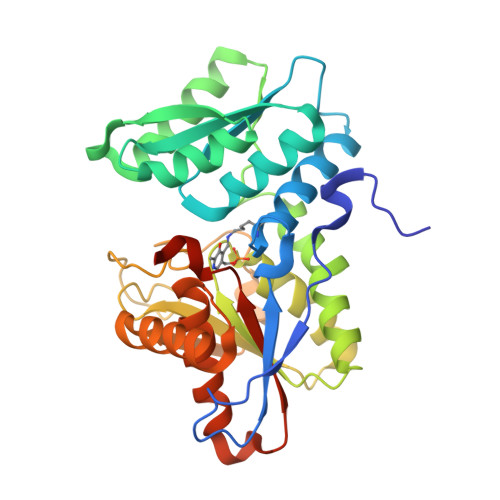
Cysteine biosynthetic genes are up-regulated in the persistent phase of Mycobacterium tuberculosis, and the corresponding enzymes are therefore of interest as potential targets for novel antibacterial agents. cysK1 is one of these genes and has been annotated as coding for an O-acetylserine sulfhydrylase. Recombinant CysK1 is a pyridoxal phosphate (PLP)-dependent enzyme that catalyzes the conversion of O-acetylserine to cysteine. The crystal structure of the enzyme was determined to 1.8A resolution. CysK1 belongs to the family of fold type II PLP enzymes and is similar in structure to other O-acetylserine sulfhydrylases. We were able to trap the alpha-aminoacrylate reaction intermediate and determine its structure by cryocrystallography. Formation of the aminoacrylate complex is accompanied by a domain rotation resulting in active site closure. The aminoacrylate moiety is bound in the active site via the covalent linkage to the PLP cofactor and by hydrogen bonds of its carboxyl group to several enzyme residues. The catalytic lysine residue is positioned such that it can protonate the Calpha-carbon atom of the aminoacrylate only from the si-face, resulting in the formation of L-cysteine. CysK1 is competitively inhibited by a four-residue peptide derived from the C-terminal of serine acetyl transferase. The crystallographic analysis reveals that the peptide binds to the enzyme active site, suggesting that CysK1 forms an bi-enzyme complex with serine acetyl transferase, in a similar manner to other bacterial and plant O-acetylserine sulfhydrylases. The structure of the enzyme-peptide complex provides a framework for the design of strong binding inhibitors.
Organizational Affiliation:
Department of Medical Biochemistry and Biophysics, Karolinska Institutet, S-171 77 Stockholm, Sweden.