2Q1F

Crystal structure of chondroitin sulfate lyase abc from bacteroides thetaiotaomicron wal2926


Experimental Data Snapshot

  • Method: X-RAY DIFFRACTION
  • Resolution: 2.85 Å
  • R-Value Free: 0.262 
  • R-Value Work: 0.220 
  • R-Value Observed: 0.222 

wwPDB Validation   3D Report Full Report


This is version 1.2 of the entry. See complete history


Literature

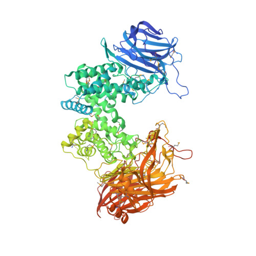
Composite active site of chondroitin lyase ABC accepting both epimers of uronic acid.

Shaya, D.Hahn, B.S.Bjerkan, T.M.Kim, W.S.Park, N.Y.Sim, J.S.Kim, Y.S.Cygler, M.

(2008) Glycobiology 18: 270-277

  • DOI: https://doi.org/10.1093/glycob/cwn002
  • Primary Citation of Related Structures:  
    2Q1F

  • PubMed Abstract: 

    Enzymes have evolved as catalysts with high degrees of stereospecificity. When both enantiomers are biologically important, enzymes with two different folds usually catalyze reactions with the individual enantiomers. In rare cases a single enzyme can process both enantiomers efficiently, but no molecular basis for such catalysis has been established. The family of bacterial chondroitin lyases ABC comprises such enzymes. They can degrade both chondroitin sulfate (CS) and dermatan sulfate (DS) glycosaminoglycans at the nonreducing end of either glucuronic acid (CS) or its epimer iduronic acid (DS) by a beta-elimination mechanism, which commences with the removal of the C-5 proton from the uronic acid. Two other structural folds evolved to perform these reactions in an epimer-specific fashion: (alpha/alpha)(5) for CS (chondroitin lyases AC) and beta-helix for DS (chondroitin lyases B); their catalytic mechanisms have been established at the molecular level. The structure of chondroitinase ABC from Proteus vulgaris showed surprising similarity to chondroitinase AC, including the presence of a Tyr-His-Glu-Arg catalytic tetrad, which provided a possible mechanism for CS degradation but not for DS degradation. We determined the structure of a distantly related Bacteroides thetaiotaomicron chondroitinase ABC to identify additional structurally conserved residues potentially involved in catalysis. We found a conserved cluster located approximately 12 A from the catalytic tetrad. We demonstrate that a histidine in this cluster is essential for catalysis of DS but not CS. The enzyme utilizes a single substrate-binding site while having two partially overlapping active sites catalyzing the respective reactions. The spatial separation of the two sets of residues suggests a substrate-induced conformational change that brings all catalytically essential residues close together.


  • Organizational Affiliation

    Department of Biochemistry, McGill University, Montréal, Québec, Canada.


Macromolecules
Find similar proteins by:  (by identity cutoff)  |  3D Structure
Entity ID: 1
MoleculeChains Sequence LengthOrganismDetailsImage
Chondroitinase
A, B
1,022Bacteroides thetaiotaomicronMutation(s): 0 
Gene Names: cslABC
EC: 4.2.2.21
UniProt
Find proteins for C5G6D7 (Bacteroides thetaiotaomicron)
Explore C5G6D7 
Go to UniProtKB:  C5G6D7
Entity Groups  
Sequence Clusters30% Identity50% Identity70% Identity90% Identity95% Identity100% Identity
UniProt GroupC5G6D7
Sequence Annotations
Expand
  • Reference Sequence
Small Molecules
Ligands 2 Unique
IDChains Name / Formula / InChI Key2D Diagram3D Interactions
PO4
Query on PO4

Download Ideal Coordinates CCD File 
D [auth A]
E [auth A]
F [auth A]
G [auth A]
H [auth A]
D [auth A],
E [auth A],
F [auth A],
G [auth A],
H [auth A],
I [auth A],
J [auth A],
K [auth A],
M [auth B],
N [auth B],
O [auth B],
P [auth B],
Q [auth B],
R [auth B],
S [auth B],
T [auth B]
PHOSPHATE ION
O4 P
NBIIXXVUZAFLBC-UHFFFAOYSA-K
CA
Query on CA

Download Ideal Coordinates CCD File 
C [auth A],
L [auth B]
CALCIUM ION
Ca
BHPQYMZQTOCNFJ-UHFFFAOYSA-N
Modified Residues  1 Unique
IDChains TypeFormula2D DiagramParent
MSE
Query on MSE
A, B
L-PEPTIDE LINKINGC5 H11 N O2 SeMET
Experimental Data & Validation

Experimental Data

  • Method: X-RAY DIFFRACTION
  • Resolution: 2.85 Å
  • R-Value Free: 0.262 
  • R-Value Work: 0.220 
  • R-Value Observed: 0.222 
  • Space Group: P 63
Unit Cell:
Length ( Å )Angle ( ˚ )
a = 223.362α = 90
b = 223.362β = 90
c = 112.639γ = 120
Software Package:
Software NamePurpose
DENZOdata reduction
SCALEPACKdata scaling
RESOLVEphasing
REFMACrefinement
PDB_EXTRACTdata extraction
HKL-2000data reduction
SHARPphasing

Structure Validation

View Full Validation Report


Entry History 

Deposition Data

Revision History  (Full details and data files)

  • Version 1.0: 2008-01-29
    Type: Initial release
  • Version 1.1: 2011-07-13
    Changes: Advisory, Version format compliance
  • Version 1.2: 2017-10-18
    Changes: Refinement description