Structural and functional analysis of the ligand specificity of the HtrA2/Omi PDZ domain.
Zhang, Y., Appleton, B.A., Wu, P., Wiesmann, C., Sidhu, S.S.(2007) Protein Sci 16: 1738-1750
- PubMed: 17656586
- DOI: https://doi.org/10.1110/ps.072833207
- Primary Citation of Related Structures:
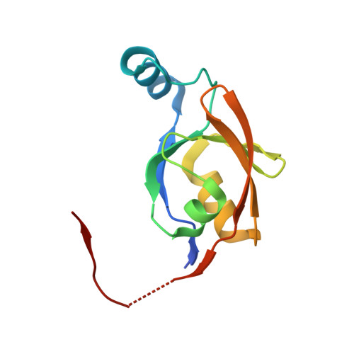
2PZD - PubMed Abstract:
The mitochondrial serine protease HtrA2/Omi helps to maintain mitochondrial function by handling misfolded proteins in the intermembrane space. In addition, HtrA2/Omi has been implicated as a proapoptotic factor upon release into the cytoplasm during the cell death cascade. The protein contains a C-terminal PDZ domain that packs against the protease active site and inhibits proteolytic activity. Engagement of the PDZ domain by peptide ligands has been shown to activate the protease and also has been proposed to mediate substrate recognition. We report a detailed structural and functional analysis of the human HtrA2/Omi PDZ domain using peptide libraries and affinity assays to define specificity, X-ray crystallography to view molecular details of PDZ-ligand interactions, and alanine-scanning mutagenesis to probe the peptide-binding groove. We show that the HtrA2/Omi PDZ domain recognizes both C-terminal and internal stretches of extended, hydrophobic polypeptides. High-affinity ligand recognition requires contacts with up to five hydrophobic side chains by distinct sites on the PDZ domain. However, no particular residue type is absolutely required at any position, and thus, the HtrA2/Omi PDZ domain appears to be a promiscuous module adapted to recognize unstructured, hydrophobic polypeptides. This type of specificity is consistent with the biological role of HtrA2/Omi in mitochondria, which requires the recognition of diverse, exposed stretches of hydrophobic sequences in misfolded proteins. The findings are less consistent with, but do not exclude, a role for the PDZ domain in targeting the protease to specific substrates during apoptosis.
Organizational Affiliation:
Department of Protein Engineering, Genentech, Inc., South San Francisco, California 94080, USA.