2PYY

Crystal Structure of the GluR0 ligand-binding core from Nostoc punctiforme in complex with (L)-glutamate


Experimental Data Snapshot

  • Method: X-RAY DIFFRACTION
  • Resolution: 2.10 Å
  • R-Value Free: 0.267 
  • R-Value Work: 0.233 
  • R-Value Observed: 0.242 

wwPDB Validation   3D Report Full Report


This is version 1.2 of the entry. See complete history


Literature

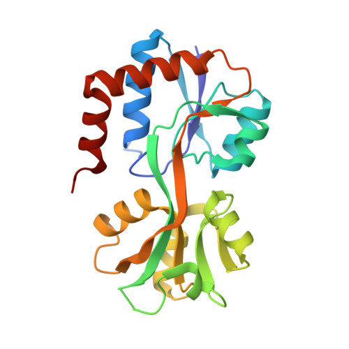
Crystal structure of the GluR0 ligand-binding core from Nostoc punctiforme in complex with L-glutamate: structural dissection of the ligand interaction and subunit interface.

Lee, J.H.Kang, G.B.Lim, H.H.Jin, K.S.Kim, S.H.Ree, M.Park, C.S.Kim, S.J.Eom, S.H.

(2008) J Mol Biol 376: 308-316

  • DOI: https://doi.org/10.1016/j.jmb.2007.10.081
  • Primary Citation of Related Structures:  
    2PYY

  • PubMed Abstract: 

    GluR0 from Nostoc punctiforme (NpGluR0) is a bacterial homologue of the ionotropic glutamate receptor (iGluR). We have solved the crystal structure of the ligand-binding core of NpGluR0 in complex with l-glutamate at a resolution of 2.1 A. The structure exhibits a noncanonical ligand interaction and two distinct subunit interfaces. The side-chain guanidium group of Arg80 forms a salt bridge with the gamma-carboxyl group of bound L-glutamate: in GluR0 from Synechocystis (SGluR0) and other iGluRs, the equivalent residues are Asn or Thr, which cannot form a similar interaction. We suggest that the local positively charged environment and the steric constraint created by Arg80 mediate the selectivity of L-glutamate binding by preventing the binding of positively charged and hydrophobic amino acids. In addition, the NpGluR0 ligand-binding core forms a new subunit interface in which the two protomers are arranged differently than the known iGluR and SGluR0 dimer interfaces. The significance of there being two different dimer interfaces was investigated using analytical ultracentrifugation analysis.


  • Organizational Affiliation

    Department of Life Science, Cell Dynamics Research Center, Gwangju Institute of Science and Technology (GIST), Gwangju 500-712, Republic of Korea.


Macromolecules
Find similar proteins by:  (by identity cutoff)  |  3D Structure
Entity ID: 1
MoleculeChains Sequence LengthOrganismDetailsImage
Ionotropic glutamate receptor bacterial homologue
A, B, C
228Nostoc punctiforme PCC 73102Mutation(s): 0 
UniProt
Find proteins for D0VWX9 (Nostoc punctiforme (strain ATCC 29133 / PCC 73102))
Explore D0VWX9 
Go to UniProtKB:  D0VWX9
Entity Groups  
Sequence Clusters30% Identity50% Identity70% Identity90% Identity95% Identity100% Identity
UniProt GroupD0VWX9
Sequence Annotations
Expand
  • Reference Sequence
Small Molecules
Binding Affinity Annotations 
IDSourceBinding Affinity
GLU Binding MOAD:  2PYY Kd: 2.30e+4 (nM) from 1 assay(s)
PDBBind:  2PYY Kd: 2.50e+4 (nM) from 1 assay(s)
Experimental Data & Validation

Experimental Data

  • Method: X-RAY DIFFRACTION
  • Resolution: 2.10 Å
  • R-Value Free: 0.267 
  • R-Value Work: 0.233 
  • R-Value Observed: 0.242 
  • Space Group: C 2 2 21
Unit Cell:
Length ( Å )Angle ( ˚ )
a = 77.979α = 90
b = 145.085β = 90
c = 132.128γ = 90
Software Package:
Software NamePurpose
HKL-2000data collection
MOLREPphasing
CNSrefinement
HKL-2000data reduction
HKL-2000data scaling

Structure Validation

View Full Validation Report


Entry History 

Deposition Data

Revision History  (Full details and data files)

  • Version 1.0: 2008-01-22
    Type: Initial release
  • Version 1.1: 2011-07-13
    Changes: Source and taxonomy, Version format compliance
  • Version 1.2: 2023-08-30
    Changes: Data collection, Database references, Derived calculations, Refinement description