2PYU

Structure of the E. coli inosine triphosphate pyrophosphatase RgdB in complex with IMP


Experimental Data Snapshot

  • Method: X-RAY DIFFRACTION
  • Resolution: 2.02 Å
  • R-Value Free: 0.253 
  • R-Value Work: 0.192 
  • R-Value Observed: 0.195 

wwPDB Validation   3D Report Full Report


Ligand Structure Quality Assessment 


This is version 1.3 of the entry. See complete history


Literature

Molecular basis of the antimutagenic activity of the house-cleaning inosine triphosphate pyrophosphatase RdgB from Escherichia coli.

Savchenko, A.Proudfoot, M.Skarina, T.Singer, A.Litvinova, O.Sanishvili, R.Brown, G.Chirgadze, N.Yakunin, A.F.

(2007) J Mol Biol 374: 1091-1103

  • DOI: https://doi.org/10.1016/j.jmb.2007.10.012
  • Primary Citation of Related Structures:  
    1K7K, 2PYU, 2Q16

  • PubMed Abstract: 

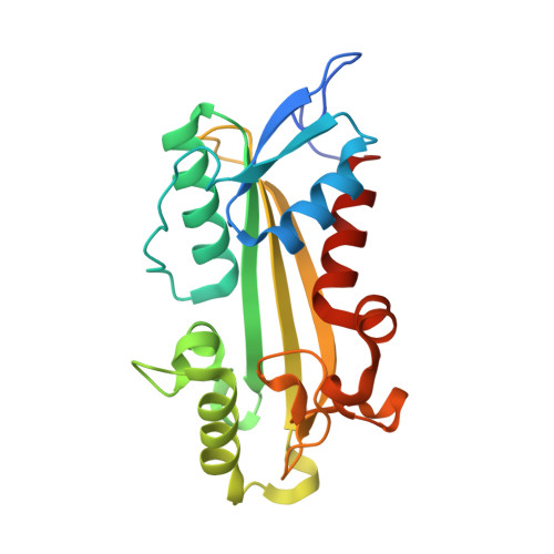
    Inosine triphosphate pyrophosphatases, which are ubiquitous house-cleaning enzymes, hydrolyze noncanonical nucleoside triphosphates (inosine triphosphate (ITP) and xanthosine triphosphate (XTP)) and prevent the incorporation of hypoxanthine or xanthine into nascent DNA or RNA. Here we present the 1.5-A-resolution crystal structure of the inosine triphosphate pyrophosphatase RdgB from Escherichia coli in a free state and in complex with a substrate (ITP+Ca(2+)) or a product (inosine monophosphate (IMP)). ITP binding to RdgB induced a large displacement of the alpha1 helix, closing the enzyme active site. This positions the conserved Lys13 close to the bridging oxygen between the alpha- and beta-phosphates of the substrate, weakening the P(alpha)-O bond. On the other side of the substrate, the conserved Asp69 is proposed to act as a base coordinating the catalytic water molecule. Our data provide insight into the molecular mechanisms of the substrate selectivity and catalysis of RdgB and other ITPases.


  • Organizational Affiliation

    Banting and Best Department of Medical Research and Ontario Center for Structural Proteomics, University of Toronto, 112 College Street, Toronto, Ontario, Canada M5G 1L6.


Macromolecules
Find similar proteins by:  (by identity cutoff)  |  3D Structure
Entity ID: 1
MoleculeChains Sequence LengthOrganismDetailsImage
Inosine Triphosphate Pyrophosphatase RdgB219Escherichia coli K-12Mutation(s): 1 
Gene Names: yggV
EC: 3.6.1.19
UniProt
Find proteins for P52061 (Escherichia coli (strain K12))
Explore P52061 
Go to UniProtKB:  P52061
Entity Groups  
Sequence Clusters30% Identity50% Identity70% Identity90% Identity95% Identity100% Identity
UniProt GroupP52061
Sequence Annotations
Expand
  • Reference Sequence
Small Molecules
Ligands 2 Unique
IDChains Name / Formula / InChI Key2D Diagram3D Interactions
IMP
Query on IMP

Download Ideal Coordinates CCD File 
B [auth A]INOSINIC ACID
C10 H13 N4 O8 P
GRSZFWQUAKGDAV-KQYNXXCUSA-N
EDO
Query on EDO

Download Ideal Coordinates CCD File 
C [auth A]1,2-ETHANEDIOL
C2 H6 O2
LYCAIKOWRPUZTN-UHFFFAOYSA-N
Experimental Data & Validation

Experimental Data

  • Method: X-RAY DIFFRACTION
  • Resolution: 2.02 Å
  • R-Value Free: 0.253 
  • R-Value Work: 0.192 
  • R-Value Observed: 0.195 
  • Space Group: P 43 21 2
Unit Cell:
Length ( Å )Angle ( ˚ )
a = 77.941α = 90
b = 77.941β = 90
c = 81.073γ = 90
Software Package:
Software NamePurpose
REFMACrefinement
HKL-2000data collection
HKL-2000data reduction
HKL-2000data scaling
AMoREphasing

Structure Validation

View Full Validation Report


Ligand Structure Quality Assessment 


Entry History 

Deposition Data

Revision History  (Full details and data files)

  • Version 1.0: 2008-03-18
    Type: Initial release
  • Version 1.1: 2011-07-13
    Changes: Source and taxonomy, Version format compliance
  • Version 1.2: 2021-10-20
    Changes: Database references, Derived calculations
  • Version 1.3: 2023-08-30
    Changes: Data collection, Refinement description