2PX0

Crystal structure of FlhF complexed with GMPPNP/Mg(2+)


Experimental Data Snapshot

  • Method: X-RAY DIFFRACTION
  • Resolution: 3.00 Å
  • R-Value Free: 0.328 
  • R-Value Work: 0.254 
  • R-Value Observed: 0.254 

wwPDB Validation   3D Report Full Report


Ligand Structure Quality Assessment 


This is version 1.2 of the entry. See complete history


Literature

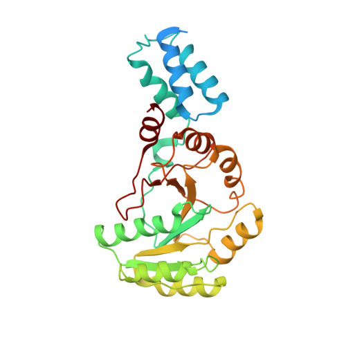
The crystal structure of the third signal-recognition particle GTPase FlhF reveals a homodimer with bound GTP.

Bange, G.Petzold, G.Wild, K.Parlitz, R.O.Sinning, I.

(2007) Proc Natl Acad Sci U S A 104: 13621-13625

  • DOI: https://doi.org/10.1073/pnas.0702570104
  • Primary Citation of Related Structures:  
    2PX0, 2PX3

  • PubMed Abstract: 

    Flagella are well characterized as the organelles of locomotion and allow bacteria to react to environmental changes. The assembly of flagella is a multistep process and relies on a complex type III export machinery located in the cytoplasmic membrane. The FlhF protein is essential for the placement and assembly of polar flagella and has been classified as a signal-recognition particle (SRP)-type GTPase. SRP GTPases appeared early in evolution and form a unique subfamily within the guanine nucleotide binding proteins with only three members: the signal sequence-binding protein SRP54, the SRP receptor FtsY, and FlhF. We report the crystal structures of FlhF from Bacillus subtilis in complex with GTP and GMPPNP. FlhF shares SRP GTPase-specific features such as the presence of an N-terminal alpha-helical domain and the I-box insertion. It forms a symmetric homodimer sequestering a composite active site that contains two head-to-tail arranged nucleotides similar to the heterodimeric SRP-targeting complex. However, significant differences to the GTPases of SRP and the SRP receptor include the formation of a stable homodimer with GTP as well as severe modifications and even the absence of motifs involved in regulation of the other two SRP GTPases. Our results provide insights into SRP GTPases and their roles in two fundamentally different protein-targeting routes that both rely on efficient protein delivery to a secretion channel.


  • Organizational Affiliation

    Heidelberg University Biochemistry Center (BZH), INF 328, 69120 Heidelberg, Germany.


Macromolecules
Find similar proteins by:  (by identity cutoff)  |  3D Structure
Entity ID: 1
MoleculeChains Sequence LengthOrganismDetailsImage
Flagellar biosynthesis protein flhF
A, B, C, D, E
A, B, C, D, E, F, G, H
296Bacillus subtilisMutation(s): 0 
Gene Names: flhF
UniProt
Find proteins for Q01960 (Bacillus subtilis (strain 168))
Explore Q01960 
Go to UniProtKB:  Q01960
Entity Groups  
Sequence Clusters30% Identity50% Identity70% Identity90% Identity95% Identity100% Identity
UniProt GroupQ01960
Sequence Annotations
Expand
  • Reference Sequence
Small Molecules
Ligands 2 Unique
IDChains Name / Formula / InChI Key2D Diagram3D Interactions
GNP
Query on GNP

Download Ideal Coordinates CCD File 
J [auth A]
L [auth B]
N [auth C]
P [auth D]
R [auth E]
J [auth A],
L [auth B],
N [auth C],
P [auth D],
R [auth E],
T [auth F],
V [auth G],
X [auth H]
PHOSPHOAMINOPHOSPHONIC ACID-GUANYLATE ESTER
C10 H17 N6 O13 P3
UQABYHGXWYXDTK-UUOKFMHZSA-N
MG
Query on MG

Download Ideal Coordinates CCD File 
I [auth A]
K [auth B]
M [auth C]
O [auth D]
Q [auth E]
I [auth A],
K [auth B],
M [auth C],
O [auth D],
Q [auth E],
S [auth F],
U [auth G],
W [auth H]
MAGNESIUM ION
Mg
JLVVSXFLKOJNIY-UHFFFAOYSA-N
Experimental Data & Validation

Experimental Data

  • Method: X-RAY DIFFRACTION
  • Resolution: 3.00 Å
  • R-Value Free: 0.328 
  • R-Value Work: 0.254 
  • R-Value Observed: 0.254 
  • Space Group: P 1 21 1
Unit Cell:
Length ( Å )Angle ( ˚ )
a = 104.649α = 90
b = 111.376β = 89.78
c = 112.089γ = 90
Software Package:
Software NamePurpose
CNSrefinement
ADSCdata collection
MOSFLMdata reduction
SCALAdata scaling
PHASERphasing

Structure Validation

View Full Validation Report


Ligand Structure Quality Assessment 


Entry History 

Deposition Data

Revision History  (Full details and data files)

  • Version 1.0: 2007-09-25
    Type: Initial release
  • Version 1.1: 2011-07-13
    Changes: Version format compliance
  • Version 1.2: 2024-02-21
    Changes: Data collection, Database references, Derived calculations