


Structure of a mitochondrial type II peroxiredoxin from Pisum sativum
Lopez-Jaramillo, F.J., Barranco-Medina, S., Lazaro, J.J., Santoyo-Gonzalez, F.To be published.
Experimental Data Snapshot
wwPDB Validation 3D Report Full Report

Entity ID: 1 | |||||
|---|---|---|---|---|---|
| Molecule | Chains | Sequence Length | Organism | Details | Image |
| Mitochondrial peroxiredoxin | 171 | Pisum sativum | Mutation(s): 0 Gene Names: prx EC: 1.11.1.15 | 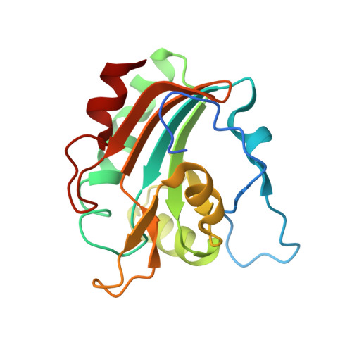 | |
UniProt | |||||
Find proteins for Q6KBB1 (Pisum sativum) Explore Q6KBB1 Go to UniProtKB: Q6KBB1 | |||||
Entity Groups | |||||
| Sequence Clusters | 30% Identity50% Identity70% Identity90% Identity95% Identity100% Identity | ||||
| UniProt Group | Q6KBB1 | ||||
Sequence AnnotationsExpand | |||||
| |||||
| Length ( Å ) | Angle ( ˚ ) |
|---|---|
| a = 61.88 | α = 102.94 |
| b = 66.4 | β = 104.44 |
| c = 77.23 | γ = 99.07 |
| Software Name | Purpose |
|---|---|
| SAINT | data scaling |
| AMoRE | phasing |
| CNS | refinement |
| PDB_EXTRACT | data extraction |
| PROTEUM PLUS | data collection |
| SAINT | data reduction |
| XPREP | data reduction |
RCSB PDB (citation) is hosted by
RCSB PDB is a member of the

