2PU9

Crystal srtucture of the binary complex between ferredoxin: thioredoxin reductase and thioredoxin f


Experimental Data Snapshot

  • Method: X-RAY DIFFRACTION
  • Resolution: 1.65 Å
  • R-Value Free: 0.231 
  • R-Value Work: 0.205 

wwPDB Validation   3D Report Full Report


This is version 1.4 of the entry. See complete history


Literature

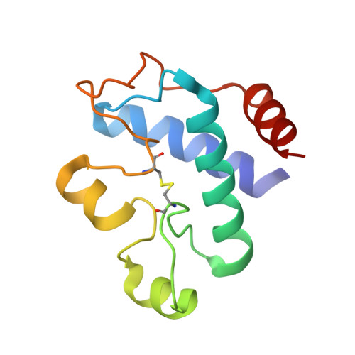
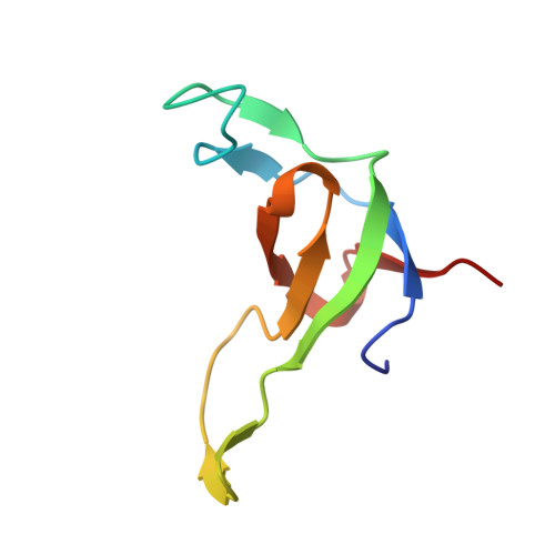
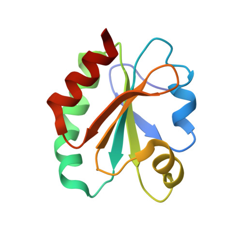
Structural snapshots along the reaction pathway of ferredoxin-thioredoxin reductase.

Dai, S.Friemann, R.Glauser, D.A.Bourquin, F.Manieri, W.Schurmann, P.Eklund, H.

(2007) Nature 448: 92-96

  • DOI: https://doi.org/10.1038/nature05937
  • Primary Citation of Related Structures:  
    2PU9, 2PUK, 2PUO, 2PVD, 2PVG, 2PVO

  • PubMed Abstract: 

    Oxygen-evolving photosynthetic organisms regulate carbon metabolism through a light-dependent redox signalling pathway. Electrons are shuttled from photosystem I by means of ferredoxin (Fdx) to ferredoxin-thioredoxin reductase (FTR), which catalyses the two-electron-reduction of chloroplast thioredoxins (Trxs). These modify target enzyme activities by reduction, regulating carbon flow. FTR is unique in its use of a [4Fe-4S] cluster and a proximal disulphide bridge in the conversion of a light signal into a thiol signal. We determined the structures of FTR in both its one- and its two-electron-reduced intermediate states and of four complexes in the pathway, including the ternary Fdx-FTR-Trx complex. Here we show that, in the first complex (Fdx-FTR) of the pathway, the Fdx [2Fe-2S] cluster is positioned suitably for electron transfer to the FTR [4Fe-4S] centre. After the transfer of one electron, an intermediate is formed in which one sulphur atom of the FTR active site is free to attack a disulphide bridge in Trx and the other sulphur atom forms a fifth ligand for an iron atom in the FTR [4Fe-4S] centre--a unique structure in biology. Fdx then delivers a second electron that cleaves the FTR-Trx heterodisulphide bond, which occurs in the Fdx-FTR-Trx complex. In this structure, the redox centres of the three proteins are aligned to maximize the efficiency of electron transfer from the Fdx [2Fe-2S] cluster to the active-site disulphide of Trxs. These results provide a structural framework for understanding the mechanism of disulphide reduction by an iron-sulphur enzyme and describe previously unknown interaction networks for both Fdx and Trx (refs 4-6).


  • Organizational Affiliation

    Howard Hughes Medical Institute, Integrated Department of Immunology, National Jewish Medical and Research Center & University of Colorado Health Sciences Center, 1400 Jackson Street, Denver, Colorado 80206, USA. dais@njc.org


Macromolecules
Find similar proteins by:  (by identity cutoff)  |  3D Structure
Entity ID: 1
MoleculeChains Sequence LengthOrganismDetailsImage
Ferredoxin-thioredoxin reductase, catalytic chain110Synechocystis sp.Mutation(s): 0 
Gene Names: ftrC
UniProt
Find proteins for Q55389 (Synechocystis sp. (strain PCC 6803 / Kazusa))
Explore Q55389 
Go to UniProtKB:  Q55389
Entity Groups  
Sequence Clusters30% Identity50% Identity70% Identity90% Identity95% Identity100% Identity
UniProt GroupQ55389
Sequence Annotations
Expand
  • Reference Sequence
Find similar proteins by:  (by identity cutoff)  |  3D Structure
Entity ID: 2
MoleculeChains Sequence LengthOrganismDetailsImage
Ferredoxin-thioredoxin reductase, variable chain74Synechocystis sp.Mutation(s): 0 
Gene Names: ftrV
UniProt
Find proteins for Q55781 (Synechocystis sp. (strain PCC 6803 / Kazusa))
Explore Q55781 
Go to UniProtKB:  Q55781
Entity Groups  
Sequence Clusters30% Identity50% Identity70% Identity90% Identity95% Identity100% Identity
UniProt GroupQ55781
Sequence Annotations
Expand
  • Reference Sequence
Find similar proteins by:  (by identity cutoff)  |  3D Structure
Entity ID: 3
MoleculeChains Sequence LengthOrganismDetailsImage
Thioredoxin F-type, chloroplast111Spinacia oleraceaMutation(s): 1 
UniProt
Find proteins for P09856 (Spinacia oleracea)
Explore P09856 
Go to UniProtKB:  P09856
Entity Groups  
Sequence Clusters30% Identity50% Identity70% Identity90% Identity95% Identity100% Identity
UniProt GroupP09856
Sequence Annotations
Expand
  • Reference Sequence
Experimental Data & Validation

Experimental Data

  • Method: X-RAY DIFFRACTION
  • Resolution: 1.65 Å
  • R-Value Free: 0.231 
  • R-Value Work: 0.205 
  • Space Group: P 21 21 21
Unit Cell:
Length ( Å )Angle ( ˚ )
a = 61.322α = 90
b = 66.788β = 90
c = 69.698γ = 90
Software Package:
Software NamePurpose
DENZOdata reduction
SCALEPACKdata scaling
AMoREphasing
CNSrefinement
PDB_EXTRACTdata extraction
HKL-2000data reduction
HKL-2000data scaling

Structure Validation

View Full Validation Report


Entry History 

Deposition Data

  • Released Date: 2007-07-10 
  • Deposition Author(s): Dai, S.

Revision History  (Full details and data files)

  • Version 1.0: 2007-07-10
    Type: Initial release
  • Version 1.1: 2008-05-01
    Changes: Version format compliance
  • Version 1.2: 2011-07-13
    Changes: Version format compliance
  • Version 1.3: 2017-10-18
    Changes: Refinement description
  • Version 1.4: 2021-10-20
    Changes: Database references, Derived calculations