Substrate and Product Complexes of Escherichia coli Adenylosuccinate Lyase Provide New Insights into the Enzymatic Mechanism.
Tsai, M., Koo, J., Yip, P., Colman, R.F., Segall, M.L., Howell, P.L.(2007) J Mol Biol 370: 541-554
- PubMed: 17531264
- DOI: https://doi.org/10.1016/j.jmb.2007.04.052
- Primary Citation of Related Structures:
2PTQ, 2PTR, 2PTS - PubMed Abstract:
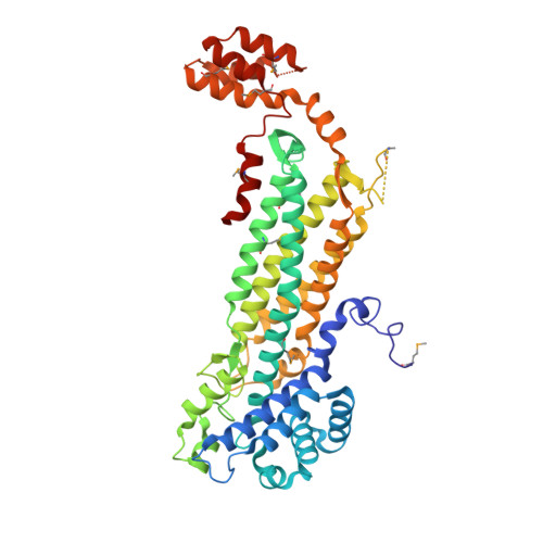
Adenylosuccinate lyase (ADL) catalyzes the breakdown of 5-aminoimidazole- (N-succinylocarboxamide) ribotide (SAICAR) to 5-aminoimidazole-4-carboxamide ribotide (AICAR) and fumarate, and of adenylosuccinate (ADS) to adenosine monophosphate (AMP) and fumarate in the de novo purine biosynthetic pathway. ADL belongs to the argininosuccinate lyase (ASL)/fumarase C superfamily of enzymes. Members of this family share several common features including: a mainly alpha-helical, homotetrameric structure; three regions of highly conserved amino acid residues; and a general acid-base catalytic mechanism with the overall beta-elimination of fumarate as a product. The crystal structures of wild-type Escherichia coli ADL (ec-ADL), and mutant-substrate (H171A-ADS) and -product (H171N-AMP.FUM) complexes have been determined to 2.0, 1.85, and 2.0 A resolution, respectively. The H171A-ADS and H171N-AMP.FUM structures provide the first detailed picture of the ADL active site, and have enabled the precise identification of substrate binding and putative catalytic residues. Contrary to previous suggestions, the ec-ADL structures implicate S295 and H171 in base and acid catalysis, respectively. Furthermore, structural alignments of ec-ADL with other superfamily members suggest for the first time a large conformational movement of the flexible C3 loop (residues 287-303) in ec-ADL upon substrate binding and catalysis, resulting in its closure over the active site. This loop movement has been observed in other superfamily enzymes, and has been proposed to be essential for catalysis. The ADL catalytic mechanism is re-examined in light of the results presented here.
Organizational Affiliation:
Molecular Structure and Function, Research Institute, Hospital for Sick Children, Toronto, Ontario, Canada M5G 1X8.