2PR5

Structural Basis for Light-dependent Signaling in the Dimeric LOV Photosensor YtvA (Dark Structure)


Experimental Data Snapshot

  • Method: X-RAY DIFFRACTION
  • Resolution: 1.45 Å
  • R-Value Free: 0.224 
  • R-Value Work: 0.190 
  • R-Value Observed: 0.192 

wwPDB Validation   3D Report Full Report


Ligand Structure Quality Assessment 


This is version 1.3 of the entry. See complete history


Literature

Structural Basis for Light-dependent Signaling in the Dimeric LOV Domain of the Photosensor YtvA.

Moglich, A.Moffat, K.

(2007) J Mol Biol 373: 112-126

  • DOI: https://doi.org/10.1016/j.jmb.2007.07.039
  • Primary Citation of Related Structures:  
    2PR5, 2PR6

  • PubMed Abstract: 

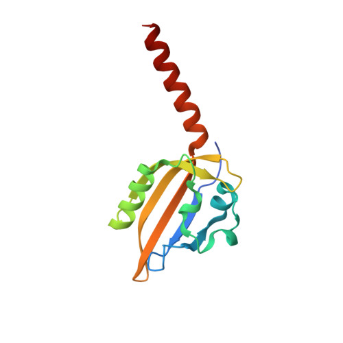
    The photosensor YtvA binds flavin mononucleotide and regulates the general stress reaction in Bacillus subtilis in response to blue light illumination. It belongs to the family of light-oxygen-voltage (LOV) proteins that were first described in plant phototropins and form a subgroup of the Per-Arnt-Sim (PAS) superfamily. Here, we report the three-dimensional structure of the LOV domain of YtvA in its dark and light states. The protein assumes the global fold common to all PAS domains and dimerizes via a hydrophobic interface. Directly C-terminal to the core of the LOV domain, an alpha-helix extends into the solvent. Light absorption causes formation of a covalent bond between a conserved cysteine residue and atom C(4a) of the FMN ring, which triggers rearrangements throughout the LOV domain. Concomitantly, in the dark and light structures, the two subunits of the dimeric protein rotate relative to each other by 5 degrees . This small quaternary structural change is presumably a component of the mechanism by which the activity of YtvA is regulated in response to light. In terms of both structure and signaling mechanism, YtvA differs from plant phototropins and more closely resembles prokaryotic heme-binding PAS domains.


  • Organizational Affiliation

    Department of Biochemistry and Molecular Biology, Institute for Biophysical Dynamics, University of Chicago, 929 East 57th Street, Chicago, IL 60637, USA.


Macromolecules
Find similar proteins by:  (by identity cutoff)  |  3D Structure
Entity ID: 1
MoleculeChains Sequence LengthOrganismDetailsImage
Blue-light photoreceptor
A, B
132Bacillus subtilisMutation(s): 0 
Gene Names: pfyP
UniProt
Find proteins for O34627 (Bacillus subtilis (strain 168))
Explore O34627 
Go to UniProtKB:  O34627
Entity Groups  
Sequence Clusters30% Identity50% Identity70% Identity90% Identity95% Identity100% Identity
UniProt GroupO34627
Sequence Annotations
Expand
  • Reference Sequence
Experimental Data & Validation

Experimental Data

  • Method: X-RAY DIFFRACTION
  • Resolution: 1.45 Å
  • R-Value Free: 0.224 
  • R-Value Work: 0.190 
  • R-Value Observed: 0.192 
  • Space Group: P 21 21 2
Unit Cell:
Length ( Å )Angle ( ˚ )
a = 88.846α = 90
b = 91.634β = 90
c = 34.239γ = 90
Software Package:
Software NamePurpose
DENZOdata reduction
SCALEPACKdata scaling
REFMACrefinement
PDB_EXTRACTdata extraction
HKL-2000data reduction
HKL-2000data scaling
PHASERphasing

Structure Validation

View Full Validation Report


Ligand Structure Quality Assessment 


Entry History 

Deposition Data

Revision History  (Full details and data files)

  • Version 1.0: 2007-08-07
    Type: Initial release
  • Version 1.1: 2011-07-13
    Changes: Advisory, Derived calculations, Version format compliance
  • Version 1.2: 2017-10-18
    Changes: Refinement description
  • Version 1.3: 2024-02-21
    Changes: Data collection, Database references, Derived calculations