


Crystal structure of Hyopthetical protein (gk_1056) from geobacillus Kaustophilus HTA426
Kanaujia, S.P., Jeyakanthan, J., Kavyashree, M., Sekar, K., Agari, Y., Ebihara, A., Kuramitsu, S., Shinkai, A., Shiro, Y., Yokoyama, S.To be published.
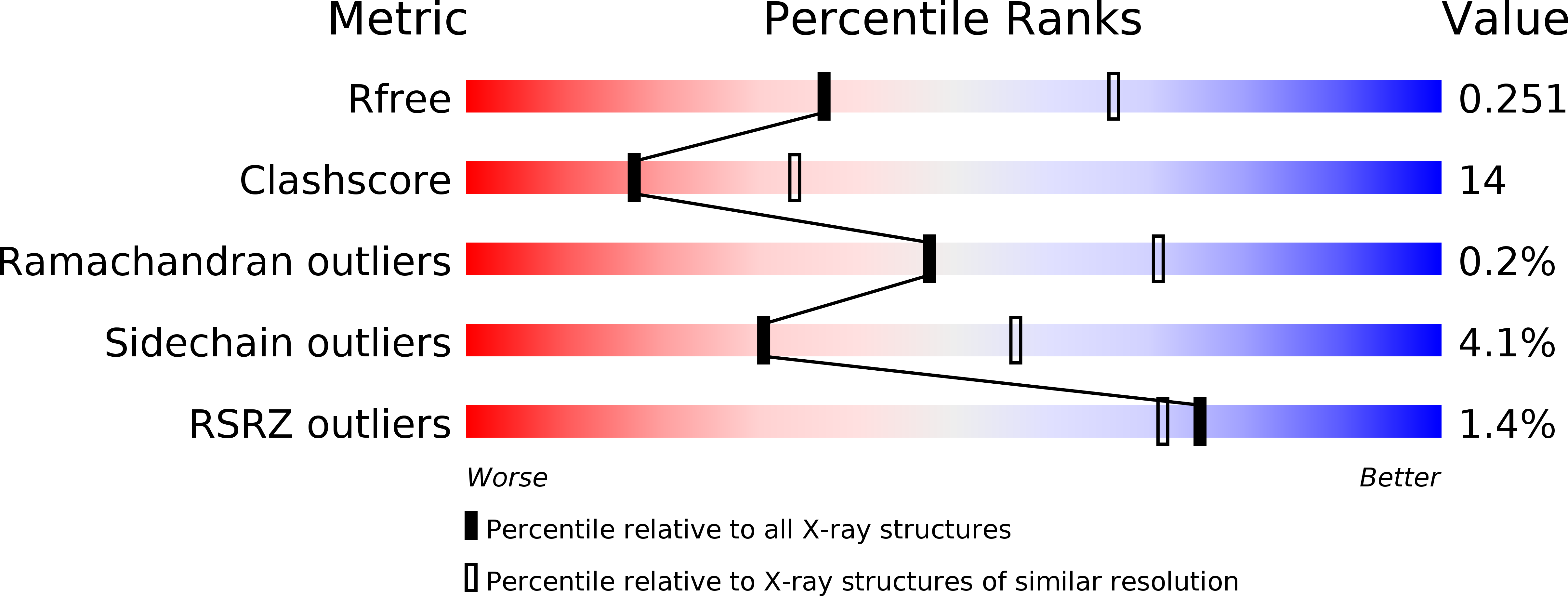
Experimental Data Snapshot
wwPDB Validation 3D Report Full Report
Entity ID: 1 | |||||
|---|---|---|---|---|---|
| Molecule | Chains | Sequence Length | Organism | Details | Image |
| Hypothetical conserved protein GK1056 | 258 | Geobacillus kaustophilus HTA426 | Mutation(s): 0 |  | |
UniProt | |||||
Find proteins for Q5L139 (Geobacillus kaustophilus (strain HTA426)) Explore Q5L139 Go to UniProtKB: Q5L139 | |||||
Entity Groups | |||||
| Sequence Clusters | 30% Identity50% Identity70% Identity90% Identity95% Identity100% Identity | ||||
| UniProt Group | Q5L139 | ||||
Sequence AnnotationsExpand | |||||
| |||||
| Modified Residues 1 Unique | |||||
|---|---|---|---|---|---|
| ID | Chains | Type | Formula | 2D Diagram | Parent |
| MSE Query on MSE | A, B | L-PEPTIDE LINKING | C5 H11 N O2 Se |  | MET |
| Length ( Å ) | Angle ( ˚ ) |
|---|---|
| a = 114.05 | α = 90 |
| b = 114.05 | β = 90 |
| c = 182.309 | γ = 90 |
| Software Name | Purpose |
|---|---|
| CNS | refinement |
| HKL-2000 | data collection |
| HKL-2000 | data reduction |
| SCALEPACK | data scaling |
| SOLVE | phasing |
RCSB PDB (citation) is hosted by
RCSB PDB is a member of the

