2PMU

Crystal structure of the DNA-binding domain of PhoP


Experimental Data Snapshot

  • Method: X-RAY DIFFRACTION
  • Resolution: 1.78 Å
  • R-Value Free: 0.238 
  • R-Value Work: 0.196 
  • R-Value Observed: 0.198 

wwPDB Validation   3D Report Full Report


This is version 1.3 of the entry. See complete history


Literature

Structure of the DNA-binding domain of the response regulator PhoP from Mycobacterium tuberculosis

Wang, S.Engohang-Ndong, J.Smith, I.

(2007) Biochemistry 46: 14751-14761

  • DOI: https://doi.org/10.1021/bi700970a
  • Primary Citation of Related Structures:  
    2PMU

  • PubMed Abstract: 

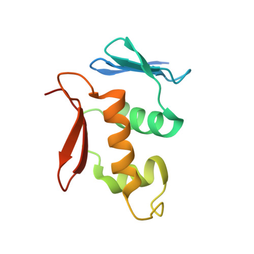
    The PhoP-PhoR two-component signaling system from Mycobacterium tuberculosis is essential for the virulence of the tubercle bacillus. The response regulator, PhoP, regulates expression of over 110 genes. In order to elucidate the regulatory mechanism of PhoP, we determined the crystal structure of its DNA-binding domain (PhoPC). PhoPC exhibits a typical fold of the winged helix-turn-helix subfamily of response regulators. The structure starts with a four-stranded antiparallel beta-sheet, followed by a three-helical bundle of alpha-helices, and then a C-terminal beta-hairpin, which together with a short beta-strand between the first and second helices forms a three-stranded antiparallel beta-sheet. Structural elements are packed through a hydrophobic core, with the first helix providing a scaffold for the rest of the domain to pack. The second and third helices and the long, flexible loop between them form the helix-turn-helix motif, with the third helix being the recognition helix. The C-terminal beta-hairpin turn forms the wing motif. The molecular surfaces around the recognition helix and the wing residues show strong positive electrostatic potential, consistent with their roles in DNA binding and nucleotide sequence recognition. The crystal packing of PhoPC gives a hexamer ring, with neighboring molecules interacting in a head-to-tail fashion. This packing interface suggests that PhoPC could bind DNA in a tandem association. However, this mode of DNA binding is likely to be nonspecific because the recognition helix is partially blocked and would be prevented from inserting into the major groove of DNA. Detailed structural analysis and implications with respect to DNA binding are discussed.


  • Organizational Affiliation

    Public Health Research Institute, New Jersey Medical School, University of Medicine and Dentistry of New Jersey, Newark, NJ 07103, USA. wang16@umdnj.edu


Macromolecules
Find similar proteins by:  (by identity cutoff)  |  3D Structure
Entity ID: 1
MoleculeChains Sequence LengthOrganismDetailsImage
response regulator PHOP
A, B, C, D, E
A, B, C, D, E, F
110Mycobacterium tuberculosis H37RvMutation(s): 0 
Gene Names: PhoP
UniProt
Find proteins for Q7D9B8 (Mycobacterium tuberculosis (strain CDC 1551 / Oshkosh))
Explore Q7D9B8 
Go to UniProtKB:  Q7D9B8
Entity Groups  
Sequence Clusters30% Identity50% Identity70% Identity90% Identity95% Identity100% Identity
UniProt GroupQ7D9B8
Sequence Annotations
Expand
  • Reference Sequence
Small Molecules
Ligands 5 Unique
IDChains Name / Formula / InChI Key2D Diagram3D Interactions
PO4
Query on PO4

Download Ideal Coordinates CCD File 
G [auth A],
O [auth D],
P [auth D],
T [auth E]
PHOSPHATE ION
O4 P
NBIIXXVUZAFLBC-UHFFFAOYSA-K
GLY
Query on GLY

Download Ideal Coordinates CCD File 
K [auth B],
L [auth B],
M [auth B]
GLYCINE
C2 H5 N O2
DHMQDGOQFOQNFH-UHFFFAOYSA-N
K
Query on K

Download Ideal Coordinates CCD File 
H [auth A],
Q [auth D],
U [auth E]
POTASSIUM ION
K
NPYPAHLBTDXSSS-UHFFFAOYSA-N
CL
Query on CL

Download Ideal Coordinates CCD File 
J [auth B],
R [auth D],
V [auth E],
W [auth E]
CHLORIDE ION
Cl
VEXZGXHMUGYJMC-UHFFFAOYSA-M
UNX
Query on UNX

Download Ideal Coordinates CCD File 
I [auth A],
N [auth B],
S [auth D],
X [auth E]
UNKNOWN ATOM OR ION
X
Experimental Data & Validation

Experimental Data

  • Method: X-RAY DIFFRACTION
  • Resolution: 1.78 Å
  • R-Value Free: 0.238 
  • R-Value Work: 0.196 
  • R-Value Observed: 0.198 
  • Space Group: C 1 2 1
Unit Cell:
Length ( Å )Angle ( ˚ )
a = 103.884α = 90
b = 101.129β = 126.72
c = 86.93γ = 90
Software Package:
Software NamePurpose
DENZOdata reduction
SCALEPACKdata scaling
REFMACrefinement
PDB_EXTRACTdata extraction
PHASERphasing

Structure Validation

View Full Validation Report


Entry History 

Deposition Data

  • Released Date: 2008-02-26 
  • Deposition Author(s): Wang, S.

Revision History  (Full details and data files)

  • Version 1.0: 2008-02-26
    Type: Initial release
  • Version 1.1: 2011-07-13
    Changes: Advisory, Source and taxonomy, Version format compliance
  • Version 1.2: 2017-10-18
    Changes: Refinement description
  • Version 1.3: 2023-08-30
    Changes: Data collection, Database references, Derived calculations, Refinement description