2PLJ

Crystal structure of lysine/ornithine decarboxylase complexed with putrescine from Vibrio vulnificus


Experimental Data Snapshot

  • Method: X-RAY DIFFRACTION
  • Resolution: 1.70 Å
  • R-Value Free: 0.213 
  • R-Value Work: 0.181 
  • R-Value Observed: 0.182 

wwPDB Validation   3D Report Full Report


Ligand Structure Quality Assessment 


This is version 1.3 of the entry. See complete history


Literature

Phylogenetic diversity and the structural basis of substrate specificity in the beta/alpha-barrel fold basic amino acid decarboxylases.

Lee, J.Michael, A.J.Martynowski, D.Goldsmith, E.J.Phillips, M.A.

(2007) J Biol Chem 282: 27115-27125

  • DOI: https://doi.org/10.1074/jbc.M704066200
  • Primary Citation of Related Structures:  
    2PLJ, 2PLK

  • PubMed Abstract: 

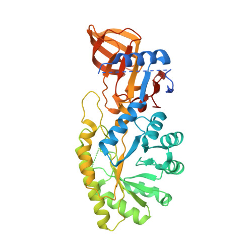
    The beta/alpha-barrel fold type basic amino acid decarboxylases include eukaryotic ornithine decarboxylases (ODC) and bacterial and plant enzymes with activity on L-arginine and meso-diaminopimelate. These enzymes catalyze essential steps in polyamine and lysine biosynthesis. Phylogenetic analysis suggests that diverse bacterial species also contain ODC-like enzymes from this fold type. However, in comparison with the eukaryotic ODCs, amino acid differences were identified in the sequence of the 3(10)-helix that forms a key specificity element in the active site, suggesting they might function on novel substrates. Putative decarboxylases from a phylogenetically diverse range of bacteria were characterized to determine their substrate preference. Enzymes from species within Methanosarcina, Pseudomonas, Bartonella, Nitrosomonas, Thermotoga, and Aquifex showed a strong preference for L-ornithine, whereas the enzyme from Vibrio vulnificus (VvL/ODC) had dual specificity functioning well on both L-ornithine and L-lysine. The x-ray structure of VvL/ODC was solved in the presence of the reaction products putrescine and cadaverine to 1.7 and 2.15A, respectively. The overall structure is similar to eukaryotic ODC; however, reorientation of the 3(10)-helix enlarging the substrate binding pocket allows L-lysine to be accommodated. The structure of the putrescine-bound enzyme suggests that a bridging water molecule between the shorter L-ornithine and key active site residues provides the structural basis for VvL/ODC to also function on this substrate. Our data demonstrate that there is greater structural and functional diversity in bacterial polyamine biosynthetic decarboxylases than previously suspected.


  • Organizational Affiliation

    Departments of Pharmacology, University of Texas Southwestern Medical Center, Dallas, Texas 75390-9041.


Macromolecules
Find similar proteins by:  (by identity cutoff)  |  3D Structure
Entity ID: 1
MoleculeChains Sequence LengthOrganismDetailsImage
lysine/ornithine decarboxylase
A, B
419Vibrio vulnificus CMCP6Mutation(s): 0 
Gene Names: LysA
EC: 4.1.1.18 (PDB Primary Data), 4.1.1.17 (PDB Primary Data)
Entity Groups  
Sequence Clusters30% Identity50% Identity70% Identity90% Identity95% Identity100% Identity
Sequence Annotations
Expand
  • Reference Sequence
Small Molecules
Ligands 2 Unique
IDChains Name / Formula / InChI Key2D Diagram3D Interactions
P3T
Query on P3T

Download Ideal Coordinates CCD File 
D [auth A],
E [auth B]
(4-{[(4-AMINOBUTYL)AMINO]METHYL}-5-HYDROXY-6-METHYLPYRIDIN-3-YL)METHYL DIHYDROGEN PHOSPHATE
C12 H22 N3 O5 P
MXDZELCJRJAUEI-UHFFFAOYSA-N
MG
Query on MG

Download Ideal Coordinates CCD File 
C [auth A]MAGNESIUM ION
Mg
JLVVSXFLKOJNIY-UHFFFAOYSA-N
Experimental Data & Validation

Experimental Data

  • Method: X-RAY DIFFRACTION
  • Resolution: 1.70 Å
  • R-Value Free: 0.213 
  • R-Value Work: 0.181 
  • R-Value Observed: 0.182 
  • Space Group: P 21 21 21
Unit Cell:
Length ( Å )Angle ( ˚ )
a = 82.074α = 90
b = 88.683β = 90
c = 111.843γ = 90
Software Package:
Software NamePurpose
REFMACrefinement
HKL-3000data collection
DENZOdata reduction
SCALEPACKdata scaling
MOLREPphasing

Structure Validation

View Full Validation Report


Ligand Structure Quality Assessment 


Entry History 

Deposition Data

Revision History  (Full details and data files)

  • Version 1.0: 2007-07-10
    Type: Initial release
  • Version 1.1: 2007-10-24
    Changes: Version format compliance
  • Version 1.2: 2011-07-13
    Changes: Advisory, Source and taxonomy, Version format compliance
  • Version 1.3: 2023-08-30
    Changes: Data collection, Database references, Derived calculations, Refinement description