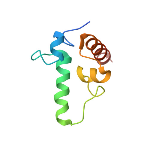
2PKG

Structure of a complex between the A subunit of protein phosphatase 2A and the small t antigen of SV40


Experimental Data Snapshot

  • Method: X-RAY DIFFRACTION
  • Resolution: 3.30 Å
  • R-Value Free: 0.312 
  • R-Value Work: 0.247 
  • R-Value Observed: 0.247 

wwPDB Validation   3D Report Full Report


This is version 1.3 of the entry. See complete history


Literature

Structural and biochemical insights into the regulation of protein phosphatase 2A by small t antigen of SV40.

Chen, Y.Xu, Y.Bao, Q.Xing, Y.Li, Z.Lin, Z.Stock, J.B.Jeffrey, P.D.Shi, Y.

(2007) Nat Struct Mol Biol 14: 527-534

  • DOI: https://doi.org/10.1038/nsmb1254

  • PubMed Abstract: 

    The small t antigen (ST) of DNA tumor virus SV40 facilitates cellular transformation by disrupting the functions of protein phosphatase 2A (PP2A) through a poorly defined mechanism. The crystal structure of the core domain of SV40 ST bound to the scaffolding subunit of human PP2A reveals that the ST core domain has a novel zinc-binding fold and interacts with the conserved ridge of HEAT repeats 3-6, which overlaps with the binding site for the B' (also called PR61 or B56) regulatory subunit. ST has a lower binding affinity than B' for the PP2A core enzyme. Consequently, ST does not efficiently displace B' from PP2A holoenzymes in vitro. Notably, ST inhibits PP2A phosphatase activity through its N-terminal J domain. These findings suggest that ST may function mainly by inhibiting the phosphatase activity of the PP2A core enzyme, and to a lesser extent by modulating assembly of the PP2A holoenzymes.


  • Organizational Affiliation

    Department of Molecular Biology, Lewis Thomas Laboratory, Princeton University, Princeton, New Jersey 08544, USA.


Macromolecules
Find similar proteins by:  (by identity cutoff)  |  3D Structure
Entity ID: 1
MoleculeChains Sequence LengthOrganismDetailsImage
Serine/threonine-protein phosphatase 2A 65 kDa regulatory subunit A alpha isoform
A, B
580Homo sapiensMutation(s): 0 
Gene Names: PPP2R1A
UniProt & NIH Common Fund Data Resources
Find proteins for P30153 (Homo sapiens)
Explore P30153 
Go to UniProtKB:  P30153
PHAROS:  P30153
GTEx:  ENSG00000105568 
Entity Groups  
Sequence Clusters30% Identity50% Identity70% Identity90% Identity95% Identity100% Identity
UniProt GroupP30153
Sequence Annotations
Expand
  • Reference Sequence
Find similar proteins by:  (by identity cutoff)  |  3D Structure
Entity ID: 2
MoleculeChains Sequence LengthOrganismDetailsImage
Small T antigen
C, D
88Betapolyomavirus macacaeMutation(s): 0 
UniProt
Find proteins for P03081 (Simian virus 40)
Explore P03081 
Go to UniProtKB:  P03081
Entity Groups  
Sequence Clusters30% Identity50% Identity70% Identity90% Identity95% Identity100% Identity
UniProt GroupP03081
Sequence Annotations
Expand
  • Reference Sequence
Experimental Data & Validation

Experimental Data

  • Method: X-RAY DIFFRACTION
  • Resolution: 3.30 Å
  • R-Value Free: 0.312 
  • R-Value Work: 0.247 
  • R-Value Observed: 0.247 
  • Space Group: C 2 2 21
Unit Cell:
Length ( Å )Angle ( ˚ )
a = 137.7α = 90
b = 147.79β = 90
c = 209.65γ = 90
Software Package:
Software NamePurpose
CNSrefinement
ADSCdata collection
DENZOdata reduction
SCALEPACKdata scaling
PHASERphasing

Structure Validation

View Full Validation Report


Entry History 

Deposition Data

Revision History  (Full details and data files)

  • Version 1.0: 2007-05-15
    Type: Initial release
  • Version 1.1: 2008-05-01
    Changes: Version format compliance
  • Version 1.2: 2011-07-13
    Changes: Version format compliance
  • Version 1.3: 2024-02-21
    Changes: Data collection, Database references, Derived calculations