2PI5

T7 RNA polymerase complexed with a phi10 promoter


Experimental Data Snapshot

  • Method: X-RAY DIFFRACTION
  • Resolution: 2.90 Å
  • R-Value Free: 0.268 
  • R-Value Work: 0.249 
  • R-Value Observed: 0.249 

wwPDB Validation   3D Report Full Report


This is version 1.3 of the entry. See complete history


Literature

Mechanism for de novo RNA synthesis and initiating nucleotide specificity by t7 RNA polymerase.

Kennedy, W.P.Momand, J.R.Yin, Y.W.

(2007) J Mol Biol 370: 256-268

  • DOI: https://doi.org/10.1016/j.jmb.2007.03.041
  • Primary Citation of Related Structures:  
    2PI4, 2PI5

  • PubMed Abstract: 

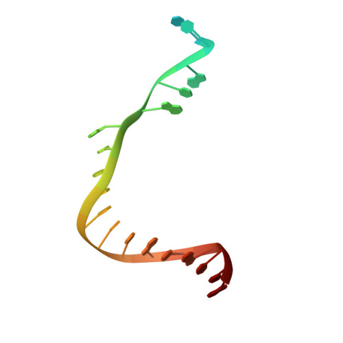
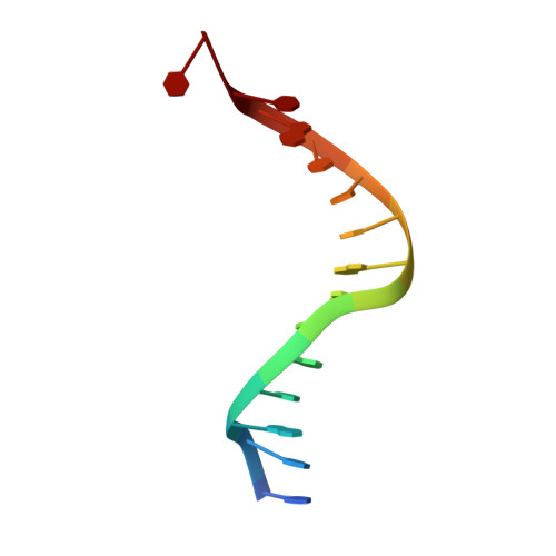
    DNA-directed RNA polymerases are capable of initiating synthesis of RNA without primers, the first catalytic stage of initiation is referred to as de novo RNA synthesis. De novo synthesis is a unique phase in the transcription cycle where the RNA polymerase binds two nucleotides rather than a nascent RNA polymer and a single nucleotide. For bacteriophage T7 RNA polymerase, transcription begins with a marked preference for GTP at the +1 and +2 positions. We determined the crystal structures of T7 RNA polymerase complexes captured during the de novo RNA synthesis. The DNA substrates in the structures in the complexes contain a common Phi 10 duplex promoter followed by a unique five base single-stranded extension of template DNA whose sequences varied at positions +1 and +2, thereby allowing for different pairs of initiating nucleotides GTP, ATP, CTP or UTP to bind. The structures show that the initiating nucleotides bind RNA polymerase in locations distinct from those described previously for elongation complexes. Selection bias in favor of GTP as an initiating nucleotide is accomplished by shape complementarity, extensive protein side-chain and strong base-stacking interactions for the guanine moiety in the enzyme active site. Consequently, an initiating GTP provides the largest stabilization force for the open promoter conformation.


  • Organizational Affiliation

    Department of Chemistry and Biochemistry, Institute of Cellular and Molecular Biology, University of Texas at Austin, Austin, TX 78712, USA.


Macromolecules

Find similar proteins by:  (by identity cutoff)  |  3D Structure
Entity ID: 3
MoleculeChains Sequence LengthOrganismDetailsImage
DNA-directed RNA polymeraseC [auth A]878Escherichia phage T7Mutation(s): 0 
EC: 2.7.7.6
UniProt
Find proteins for P00573 (Escherichia phage T7)
Explore P00573 
Go to UniProtKB:  P00573
Entity Groups  
Sequence Clusters30% Identity50% Identity70% Identity90% Identity95% Identity100% Identity
UniProt GroupP00573
Sequence Annotations
Expand
  • Reference Sequence

Find similar nucleic acids by:  Sequence   |   3D Structure  

Entity ID: 1
MoleculeChains LengthOrganismImage
5'-D(*CP*TP*TP*C*CP*TP*AP*TP*AP*GP*TP*GP*AP*GP*TP*CP*GP*TP*AP*TP*TP*A)-3'A [auth T]22N/A
Sequence Annotations
Expand
  • Reference Sequence

Find similar nucleic acids by:  Sequence   |   3D Structure  

Entity ID: 2
MoleculeChains LengthOrganismImage
5'-D(*TP*AP*AP*TP*AP*CP*GP*AP*CP*TP*CP*AP*CP*T)-3'B [auth P]14N/A
Sequence Annotations
Expand
  • Reference Sequence
Experimental Data & Validation

Experimental Data

  • Method: X-RAY DIFFRACTION
  • Resolution: 2.90 Å
  • R-Value Free: 0.268 
  • R-Value Work: 0.249 
  • R-Value Observed: 0.249 
  • Space Group: P 21 21 2
Unit Cell:
Length ( Å )Angle ( ˚ )
a = 224.45α = 90
b = 73.791β = 90
c = 80.315γ = 90
Software Package:
Software NamePurpose
CNSrefinement
DENZOdata reduction
SCALEPACKdata scaling
CNSphasing

Structure Validation

View Full Validation Report


Entry History 

Deposition Data

Revision History  (Full details and data files)

  • Version 1.0: 2007-06-19
    Type: Initial release
  • Version 1.1: 2008-05-01
    Changes: Version format compliance
  • Version 1.2: 2011-07-13
    Changes: Version format compliance
  • Version 1.3: 2024-02-21
    Changes: Data collection, Database references