2PGB

Inhibitor-free human thrombin mutant C191A-C220A


Experimental Data Snapshot

  • Method: X-RAY DIFFRACTION
  • Resolution: 1.54 Å
  • R-Value Free: 0.217 
  • R-Value Work: 0.183 
  • R-Value Observed: 0.183 

wwPDB Validation   3D Report Full Report


Ligand Structure Quality Assessment 


This is version 1.5 of the entry. See complete history


Literature

Important role of the cys-191 cys-220 disulfide bond in thrombin function and allostery

Bush-Pelc, L.A.Marino, F.Chen, Z.Pineda, A.O.Mathews, F.S.Di Cera, E.

(2007) J Biol Chem 282: 27165-27170

  • DOI: https://doi.org/10.1074/jbc.M703202200
  • Primary Citation of Related Structures:  
    2PGB, 2PGQ

  • PubMed Abstract: 

    Little is known on the role of disulfide bonds in the catalytic domain of serine proteases. The Cys-191-Cys-220 disulfide bond is located between the 190 strand leading to the oxyanion hole and the 220-loop that contributes to the architecture of the primary specificity pocket and the Na+ binding site in allosteric proteases. Removal of this bond in thrombin produces an approximately 100-fold loss of activity toward several chromogenic and natural substrates carrying Arg or Lys at P1. Na+ activation is compromised, and no fluorescence change can be detected in response to Na+ binding. A 1.54-A resolution structure of the C191A/C220A mutant in the free form reveals a conformation similar to the Na+-free slow form of wild type. The lack of disulfide bond exposes the side chain of Asp-189 to solvent, flips the backbone O atom of Gly-219, and generates disorder in portions of the 186 and 220 loops defining the Na+ site. This conformation, featuring perturbation of the Na+ site but with the active site accessible to substrate, offers a possible representation of the recently identified E* form of thrombin. Disorder in the 186 and 220 loops and the flip of Gly-219 are corrected by the active site inhibitor H-D-Phe-Pro-Arg-CH(2)Cl, as revealed by the 1.8-A resolution structure of the complex. We conclude that the Cys-191-Cys-220 disulfide bond confers stability to the primary specificity pocket by shielding Asp-189 from the solvent and orients the backbone O atom of Gly-219 for optimal substrate binding. In addition, the disulfide bond stabilizes the 186 and 220 loops that are critical for Na+ binding and activation.


  • Organizational Affiliation

    Department of Biochemistry and Molecular Biophysics, Washington University School of Medicine, St. Louis, Missouri 63110.


Macromolecules
Find similar proteins by:  (by identity cutoff)  |  3D Structure
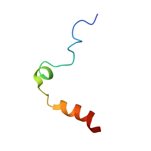
Entity ID: 1
MoleculeChains Sequence LengthOrganismDetailsImage
Prothrombin36Homo sapiensMutation(s): 0 
Gene Names: F2
EC: 3.4.21.5
UniProt & NIH Common Fund Data Resources
Find proteins for P00734 (Homo sapiens)
Explore P00734 
Go to UniProtKB:  P00734
PHAROS:  P00734
GTEx:  ENSG00000180210 
Entity Groups  
Sequence Clusters30% Identity50% Identity70% Identity90% Identity95% Identity100% Identity
UniProt GroupP00734
Sequence Annotations
Expand
  • Reference Sequence
Find similar proteins by:  (by identity cutoff)  |  3D Structure
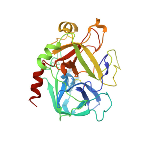
Entity ID: 2
MoleculeChains Sequence LengthOrganismDetailsImage
Prothrombin259Homo sapiensMutation(s): 2 
Gene Names: F2
EC: 3.4.21.5
UniProt & NIH Common Fund Data Resources
Find proteins for P00734 (Homo sapiens)
Explore P00734 
Go to UniProtKB:  P00734
PHAROS:  P00734
GTEx:  ENSG00000180210 
Entity Groups  
Sequence Clusters30% Identity50% Identity70% Identity90% Identity95% Identity100% Identity
UniProt GroupP00734
Sequence Annotations
Expand
  • Reference Sequence
Experimental Data & Validation

Experimental Data

  • Method: X-RAY DIFFRACTION
  • Resolution: 1.54 Å
  • R-Value Free: 0.217 
  • R-Value Work: 0.183 
  • R-Value Observed: 0.183 
  • Space Group: P 21 21 2
Unit Cell:
Length ( Å )Angle ( ˚ )
a = 61.412α = 90
b = 96.597β = 90
c = 48.14γ = 90
Software Package:
Software NamePurpose
CNSrefinement
ADSCdata collection
HKL-2000data reduction
SCALEPACKdata scaling
CNSphasing

Structure Validation

View Full Validation Report


Ligand Structure Quality Assessment 


Entry History 

Deposition Data

Revision History  (Full details and data files)

  • Version 1.0: 2007-07-17
    Type: Initial release
  • Version 1.1: 2008-04-21
    Changes: Version format compliance
  • Version 1.2: 2011-07-13
    Changes: Non-polymer description, Version format compliance
  • Version 1.3: 2020-07-29
    Type: Remediation
    Reason: Carbohydrate remediation
    Changes: Data collection, Derived calculations, Structure summary
  • Version 1.4: 2021-10-20
    Changes: Database references, Structure summary
  • Version 1.5: 2023-08-30
    Changes: Data collection, Refinement description