2P7V

Crystal structure of the Escherichia coli regulator of sigma 70, Rsd, in complex with sigma 70 domain 4


Experimental Data Snapshot

  • Method: X-RAY DIFFRACTION
  • Resolution: 2.60 Å
  • R-Value Free: 0.273 
  • R-Value Work: 0.239 
  • R-Value Observed: 0.240 

wwPDB Validation   3D Report Full Report


This is version 1.2 of the entry. See complete history


Literature

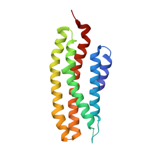
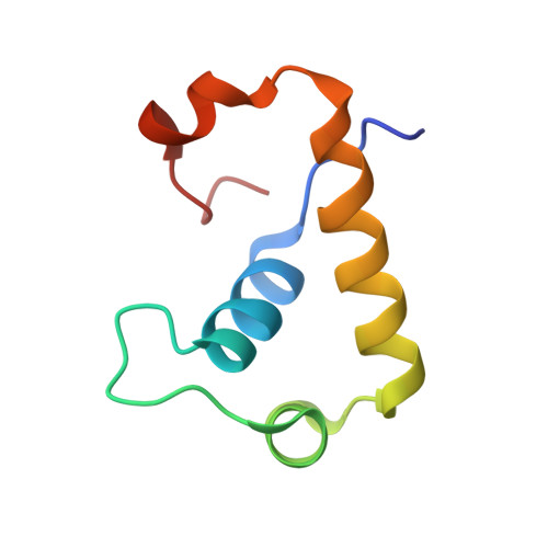
Crystal structure of the Escherichia coli regulator of sigma70, Rsd, in complex with sigma70 domain 4.

Patikoglou, G.A.Westblade, L.F.Campbell, E.A.Lamour, V.Lane, W.J.Darst, S.A.

(2007) J Mol Biol 372: 649-659

  • DOI: https://doi.org/10.1016/j.jmb.2007.06.081
  • Primary Citation of Related Structures:  
    2P7V

  • PubMed Abstract: 

    The Escherichia coli Rsd protein binds tightly and specifically to the RNA polymerase (RNAP) sigma(70) factor. Rsd plays a role in alternative sigma factor-dependent transcription by biasing the competition between sigma(70) and alternative sigma factors for the available core RNAP. Here, we determined the 2.6 A-resolution X-ray crystal structure of Rsd bound to sigma(70) domain 4 (sigma(70)(4)), the primary determinant for Rsd binding within sigma(70). The structure reveals that Rsd binding interferes with the two primary functions of sigma(70)(4), core RNAP binding and promoter -35 element binding. Interestingly, the most highly conserved Rsd residues form a network of interactions through the middle of the Rsd structure that connect the sigma(70)(4)-binding surface with three cavities exposed on distant surfaces of Rsd, suggesting functional coupling between sigma(70)(4) binding and other binding surfaces of Rsd, either for other proteins or for as yet unknown small molecule effectors. These results provide a structural basis for understanding the role of Rsd, as well as its ortholog, AlgQ, a positive regulator of Pseudomonas aeruginosa virulence, in transcription regulation.


  • Organizational Affiliation

    The Rockefeller University, Box 224, 1230 York Avenue, New York, NY 10065, USA.


Macromolecules
Find similar proteins by:  (by identity cutoff)  |  3D Structure
Entity ID: 1
MoleculeChains Sequence LengthOrganismDetailsImage
Regulator of sigma D158Escherichia coliMutation(s): 0 
Gene Names: rsd
UniProt
Find proteins for P0AFX4 (Escherichia coli (strain K12))
Explore P0AFX4 
Go to UniProtKB:  P0AFX4
Entity Groups  
Sequence Clusters30% Identity50% Identity70% Identity90% Identity95% Identity100% Identity
UniProt GroupP0AFX4
Sequence Annotations
Expand
  • Reference Sequence
Find similar proteins by:  (by identity cutoff)  |  3D Structure
Entity ID: 2
MoleculeChains Sequence LengthOrganismDetailsImage
RNA polymerase sigma factor rpoD68Escherichia coliMutation(s): 0 
Gene Names: rpoDalt
UniProt
Find proteins for Q0P6L9 (Escherichia coli)
Explore Q0P6L9 
Go to UniProtKB:  Q0P6L9
Entity Groups  
Sequence Clusters30% Identity50% Identity70% Identity90% Identity95% Identity100% Identity
UniProt GroupQ0P6L9
Sequence Annotations
Expand
  • Reference Sequence
Small Molecules
Ligands 1 Unique
IDChains Name / Formula / InChI Key2D Diagram3D Interactions
MG
Query on MG

Download Ideal Coordinates CCD File 
C [auth B]MAGNESIUM ION
Mg
JLVVSXFLKOJNIY-UHFFFAOYSA-N
Experimental Data & Validation

Experimental Data

  • Method: X-RAY DIFFRACTION
  • Resolution: 2.60 Å
  • R-Value Free: 0.273 
  • R-Value Work: 0.239 
  • R-Value Observed: 0.240 
  • Space Group: P 64
Unit Cell:
Length ( Å )Angle ( ˚ )
a = 84.111α = 90
b = 84.111β = 90
c = 84.219γ = 120
Software Package:
Software NamePurpose
CNSrefinement
PDB_EXTRACTdata extraction
HKL-2000data collection
HKL-2000data reduction
SCALEPACKdata scaling
SHARPphasing

Structure Validation

View Full Validation Report


Entry History 

Deposition Data

Revision History  (Full details and data files)

  • Version 1.0: 2007-08-21
    Type: Initial release
  • Version 1.1: 2011-07-13
    Changes: Derived calculations, Version format compliance
  • Version 1.2: 2024-02-21
    Changes: Data collection, Database references, Derived calculations