2P5O

Crystal structure of RB69 GP43 in complex with DNA containing an abasic site analog


Experimental Data Snapshot

  • Method: X-RAY DIFFRACTION
  • Resolution: 2.80 Å
  • R-Value Free: 0.290 
  • R-Value Work: 0.245 

wwPDB Validation   3D Report Full Report


This is version 1.4 of the entry. See complete history


Literature

Crystallographic snapshots of a replicative DNA polymerase encountering an abasic site.

Hogg, M.Wallace, S.S.Doublie, S.

(2004) EMBO J 23: 1483-1493

  • DOI: https://doi.org/10.1038/sj.emboj.7600150
  • Primary Citation of Related Structures:  
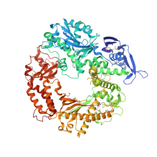
    2P5O

  • PubMed Abstract: 

    Abasic sites are common DNA lesions, which are strong blocks to replicative polymerases and are potentially mutagenic when bypassed. We report here the 2.8 A structure of the bacteriophage RB69 replicative DNA polymerase attempting to process an abasic site analog. Four different complexes were captured in the crystal asymmetric unit: two have DNA in the polymerase active site whereas the other two molecules are in the exonuclease mode. When compared to complexes with undamaged DNA, the DNA surrounding the abasic site reveals distinct changes suggesting why the lesion is so poorly bypassed: the DNA in the polymerase active site has not translocated and is therefore stalled, precluding extension. All four molecules exhibit conformations that differ from the previously published structures. The polymerase incorporates dAMP across the lesion under crystallization conditions, indicating that the different conformations observed in the crystal may be part of the active site switching reaction pathway.


  • Organizational Affiliation

    Department of Microbiology and Molecular Genetics, Markey Center for Molecular Genetics, University of Vermont, Burlington, VT, USA.


Macromolecules

Find similar proteins by:  (by identity cutoff)  |  3D Structure
Entity ID: 3
MoleculeChains Sequence LengthOrganismDetailsImage
DNA polymeraseI [auth A],
J [auth B],
K [auth C],
L [auth D]
903Escherichia phage RB69Mutation(s): 27 
Gene Names: 43
EC: 2.7.7.7
UniProt
Find proteins for Q38087 (Escherichia phage RB69)
Explore Q38087 
Go to UniProtKB:  Q38087
Entity Groups  
Sequence Clusters30% Identity50% Identity70% Identity90% Identity95% Identity100% Identity
UniProt GroupQ38087
Sequence Annotations
Expand
  • Reference Sequence

Find similar nucleic acids by:  Sequence   |   3D Structure  

Entity ID: 1
MoleculeChains LengthOrganismImage
Template DNAA [auth E],
C [auth G],
E [auth I],
G [auth K]
18N/A
Sequence Annotations
Expand
  • Reference Sequence

Find similar nucleic acids by:  Sequence   |   3D Structure  

Entity ID: 2
MoleculeChains LengthOrganismImage
Primer DNAB [auth F],
D [auth H],
F [auth J],
H [auth L]
15N/A
Sequence Annotations
Expand
  • Reference Sequence
Small Molecules
Modified Residues  1 Unique
IDChains TypeFormula2D DiagramParent
MSE
Query on MSE
I [auth A],
J [auth B],
K [auth C],
L [auth D]
L-PEPTIDE LINKINGC5 H11 N O2 SeMET
Experimental Data & Validation

Experimental Data

  • Method: X-RAY DIFFRACTION
  • Resolution: 2.80 Å
  • R-Value Free: 0.290 
  • R-Value Work: 0.245 
  • Space Group: P 1 21 1
Unit Cell:
Length ( Å )Angle ( ˚ )
a = 133.018α = 90
b = 123.209β = 95.82
c = 165.616γ = 90
Software Package:
Software NamePurpose
CBASSdata collection
CNSrefinement
DENZOdata reduction
SCALEPACKdata scaling
CNSphasing

Structure Validation

View Full Validation Report


Entry History 

Deposition Data

Revision History  (Full details and data files)

  • Version 1.0: 2007-04-03
    Type: Initial release
  • Version 1.1: 2008-05-01
    Changes: Version format compliance
  • Version 1.2: 2011-07-13
    Changes: Version format compliance
  • Version 1.3: 2014-09-10
    Changes: Database references
  • Version 1.4: 2021-10-20
    Changes: Database references, Derived calculations