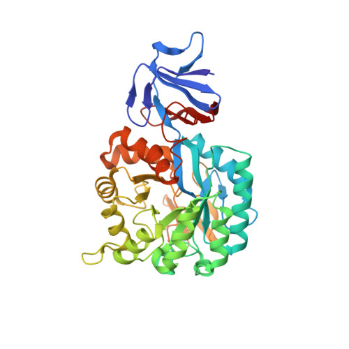
Structural Diversity within the Mononuclear and Binuclear Active Sites of N-Acetyl-d-glucosamine-6-phosphate Deacetylase.
Hall, R.S., Brown, S., Fedorov, A.A., Fedorov, E.V., Xu, C., Babbitt, P.C., Almo, S.C., Raushel, F.M.(2007) Biochemistry 46: 7953-7962
- PubMed: 17567048
- DOI: https://doi.org/10.1021/bi700544c
- Primary Citation of Related Structures:
2P50, 2P53 - PubMed Abstract:
NagA catalyzes the hydrolysis of N-acetyl-d-glucosamine-6-phosphate to d-glucosamine-6-phosphate and acetate. X-ray crystal structures of NagA from Escherichia coli were determined to establish the number and ligation scheme for the binding of zinc to the active site and to elucidate the molecular interactions between the protein and substrate. The three-dimensional structures of the apo-NagA, Zn-NagA, and the D273N mutant enzyme in the presence of a tight-binding N-methylhydroxyphosphinyl-d-glucosamine-6-phosphate inhibitor were determined. The structure of the Zn-NagA confirms that this enzyme binds a single divalent cation at the beta-position in the active site via ligation to Glu-131, His-195, and His-216. A water molecule completes the ligation shell, which is also in position to be hydrogen bonded to Asp-273. In the structure of NagA bound to the tight binding inhibitor that mimics the tetrahedral intermediate, the methyl phosphonate moiety has displaced the hydrolytic water molecule and is directly coordinated to the zinc within the active site. The side chain of Asp-273 is positioned to activate the hydrolytic water molecule via general base catalysis and to deliver this proton to the amino group upon cleavage of the amide bond of the substrate. His-143 is positioned to help polarize the carbonyl group of the substrate in conjunction with Lewis acid catalysis by the bound zinc. The inhibitor is bound in the alpha-configuration at the anomeric carbon through a hydrogen bonding interaction of the hydroxyl group at C-1 with the side chain of His-251. The phosphate group of the inhibitor attached to the hydroxyl at C-6 is ion paired with Arg-227 from the adjacent subunit. NagA from Thermotoga maritima was shown to require a single divalent cation for full catalytic activity.
Organizational Affiliation:
Department of Chemistry, P.O. Box 30012, Texas A&M University, College Station, Texas 77842-3012, USA.