2P2A

X-ray structure of the GluR2 ligand binding core (S1S2J) in complex with 2-Bn-tet-AMPA at 2.26A resolution


Experimental Data Snapshot

  • Method: X-RAY DIFFRACTION
  • Resolution: 2.26 Å
  • R-Value Free: 0.273 
  • R-Value Work: 0.211 

wwPDB Validation   3D Report Full Report


Ligand Structure Quality Assessment 


This is version 1.5 of the entry. See complete history


Literature

A tetrazolyl-substituted subtype-selective AMPA receptor agonist.

Vogensen, S.B.Frydenvang, K.Greenwood, J.R.Postorino, G.Nielsen, B.Pickering, D.S.Ebert, B.Bolcho, U.Egebjerg, J.Gajhede, M.Kastrup, J.S.Johansen, T.N.Clausen, R.P.Krogsgaard-Larsen, P.

(2007) J Med Chem 50: 2408-2414

  • DOI: https://doi.org/10.1021/jm061439q
  • Primary Citation of Related Structures:  
    2P2A

  • PubMed Abstract: 

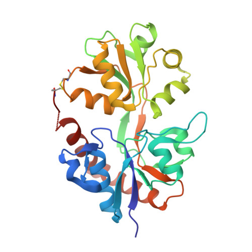
    Replacement of the methyl group of the AMPA receptor agonist 2-amino-3-[3-hydroxy-5-(2-methyl-2H-5-tetrazolyl)-4-isoxazolyl]propionic acid (2-Me-Tet-AMPA) with a benzyl group provided the first AMPA receptor agonist, compound 7, capable of discriminating GluR2-4 from GluR1 by its more than 10-fold preference for the former receptor subtypes. An X-ray crystallographic analysis of this new analogue in complex with the GluR2-S1S2J construct shows that accommodation of the benzyl group creates a previously unobserved pocket in the receptor, which may explain the remarkable pharmacological profile of compound 7.


  • Organizational Affiliation

    Department of Medicinal Chemistry, The Faculty of Pharmaceutical Sciences, University of Copenhagen, 2 Universitetsparken, DK-2100 Copenhagen, Denmark.


Macromolecules
Find similar proteins by:  (by identity cutoff)  |  3D Structure
Entity ID: 1
MoleculeChains Sequence LengthOrganismDetailsImage
Glutamate receptor 2
A, B
263Rattus norvegicusMutation(s): 0 
Gene Names: Gria2
UniProt
Find proteins for P19491 (Rattus norvegicus)
Explore P19491 
Go to UniProtKB:  P19491
Entity Groups  
Sequence Clusters30% Identity50% Identity70% Identity90% Identity95% Identity100% Identity
UniProt GroupP19491
Sequence Annotations
Expand
  • Reference Sequence
Small Molecules
Binding Affinity Annotations 
IDSourceBinding Affinity
MP9 Binding MOAD:  2P2A Ki: 750 (nM) from 1 assay(s)
PDBBind:  2P2A Ki: 750 (nM) from 1 assay(s)
Experimental Data & Validation

Experimental Data

  • Method: X-RAY DIFFRACTION
  • Resolution: 2.26 Å
  • R-Value Free: 0.273 
  • R-Value Work: 0.211 
  • Space Group: P 21 21 2
Unit Cell:
Length ( Å )Angle ( ˚ )
a = 98.12α = 90
b = 121.98β = 90
c = 47.2γ = 90
Software Package:
Software NamePurpose
CNSrefinement
PDB_EXTRACTdata extraction
MOSFLMdata reduction
SCALAdata scaling
AMoREphasing

Structure Validation

View Full Validation Report


Ligand Structure Quality Assessment 


Entry History 

Deposition Data

Revision History  (Full details and data files)

  • Version 1.0: 2007-06-19
    Type: Initial release
  • Version 1.1: 2008-05-01
    Changes: Version format compliance
  • Version 1.2: 2011-07-13
    Changes: Version format compliance
  • Version 1.3: 2017-08-16
    Changes: Data collection, Refinement description, Source and taxonomy
  • Version 1.4: 2022-12-21
    Changes: Database references, Derived calculations
  • Version 1.5: 2023-09-20
    Changes: Data collection, Refinement description