


Crystal structure of hypothetical protein (NP_085906.1) from Mesorhizobium loti at 2.15 A resolution
Joint Center for Structural Genomics (JCSG)To be published.
Experimental Data Snapshot
wwPDB Validation 3D Report Full Report

Entity ID: 1 | |||||
|---|---|---|---|---|---|
| Molecule | Chains | Sequence Length | Organism | Details | Image |
| Mll9387 protein | 286 | Mesorhizobium japonicum MAFF 303099 | Mutation(s): 11 Gene Names: NP_085906.1, mll9387 | 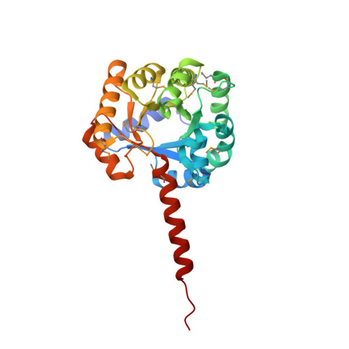 | |
UniProt | |||||
Find proteins for Q981G2 (Mesorhizobium japonicum (strain LMG 29417 / CECT 9101 / MAFF 303099)) Explore Q981G2 Go to UniProtKB: Q981G2 | |||||
Entity Groups | |||||
| Sequence Clusters | 30% Identity50% Identity70% Identity90% Identity95% Identity100% Identity | ||||
| UniProt Group | Q981G2 | ||||
Sequence AnnotationsExpand | |||||
| |||||
| Ligands 3 Unique | |||||
|---|---|---|---|---|---|
| ID | Chains | Name / Formula / InChI Key | 2D Diagram | 3D Interactions | |
| GOL Query on GOL | L [auth A], M [auth A], Q [auth B], R [auth B] | GLYCEROL C3 H8 O3 PEDCQBHIVMGVHV-UHFFFAOYSA-N |  | ||
| ACT Query on ACT | BA [auth E] CA [auth F] G [auth A] H [auth A] I [auth A] | ACETATE ION C2 H3 O2 QTBSBXVTEAMEQO-UHFFFAOYSA-M |  | ||
| CL Query on CL | AA [auth D] J [auth A] K [auth A] P [auth B] W [auth C] | CHLORIDE ION Cl VEXZGXHMUGYJMC-UHFFFAOYSA-M |  | ||
| Modified Residues 1 Unique | |||||
|---|---|---|---|---|---|
| ID | Chains | Type | Formula | 2D Diagram | Parent |
| MSE Query on MSE | A, B, C, D, E A, B, C, D, E, F | L-PEPTIDE LINKING | C5 H11 N O2 Se |  | MET |
| Length ( Å ) | Angle ( ˚ ) |
|---|---|
| a = 180.269 | α = 90 |
| b = 180.269 | β = 90 |
| c = 185.514 | γ = 120 |
| Software Name | Purpose |
|---|---|
| MolProbity | model building |
| SHELX | phasing |
| REFMAC | refinement |
| XSCALE | data scaling |
| PDB_EXTRACT | data extraction |
| XDS | data reduction |
| SHELXD | phasing |
| autoSHARP | phasing |
RCSB PDB (citation) is hosted by
RCSB PDB is a member of the

