2OZ6

Crystal Structure of Virulence Factor Regulator from Pseudomonas aeruginosa in complex with cAMP


Experimental Data Snapshot

  • Method: X-RAY DIFFRACTION
  • Resolution: 2.80 Å
  • R-Value Free: 0.251 
  • R-Value Work: 0.190 

wwPDB Validation   3D Report Full Report


Ligand Structure Quality Assessment 


This is version 1.4 of the entry. See complete history


Literature

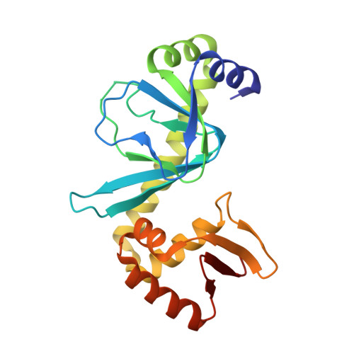
Crystal Structure of the Pseudomonas aeruginosa Virulence Factor Regulator.

Cordes, T.J.Worzalla, G.A.Ginster, A.M.Forest, K.T.

(2011) J Bacteriol 193: 4069-4074

  • DOI: https://doi.org/10.1128/JB.00666-10
  • Primary Citation of Related Structures:  
    2OZ6

  • PubMed Abstract: 

    Virulence factor regulator (Vfr) enhances Pseudomonas aeruginosa pathogenicity through its role as a global transcriptional regulator. The crystal structure of Vfr shows that it is a winged-helix DNA-binding protein like its homologue cyclic AMP receptor protein (CRP). In addition to an expected primary cyclic AMP-binding site, a second ligand-binding site is nestled between the N-terminal domain and the C-terminal helix-turn-helix domain. Unlike CRP, Vfr is a symmetric dimer in the absence of DNA. Removal of seven disordered N-terminal residues of Vfr prevents the growth of P. aeruginosa.


  • Organizational Affiliation

    University of Wisconsin-Madison, Department of Bacteriology, 1550 Linden Dr., Madison, WI 53706, USA.


Macromolecules
Find similar proteins by:  (by identity cutoff)  |  3D Structure
Entity ID: 1
MoleculeChains Sequence LengthOrganismDetailsImage
Virulence Factor Regulator207Pseudomonas aeruginosaMutation(s): 0 
Gene Names: vfr
UniProt
Find proteins for P55222 (Pseudomonas aeruginosa (strain ATCC 15692 / DSM 22644 / CIP 104116 / JCM 14847 / LMG 12228 / 1C / PRS 101 / PAO1))
Explore P55222 
Go to UniProtKB:  P55222
Entity Groups  
Sequence Clusters30% Identity50% Identity70% Identity90% Identity95% Identity100% Identity
UniProt GroupP55222
Sequence Annotations
Expand
  • Reference Sequence
Small Molecules
Ligands 1 Unique
IDChains Name / Formula / InChI Key2D Diagram3D Interactions
CMP
Query on CMP

Download Ideal Coordinates CCD File 
B [auth A],
C [auth A]
ADENOSINE-3',5'-CYCLIC-MONOPHOSPHATE
C10 H12 N5 O6 P
IVOMOUWHDPKRLL-KQYNXXCUSA-N
Binding Affinity Annotations 
IDSourceBinding Affinity
CMP PDBBind:  2OZ6 Kd: 5500 (nM) from 1 assay(s)
Experimental Data & Validation

Experimental Data

  • Method: X-RAY DIFFRACTION
  • Resolution: 2.80 Å
  • R-Value Free: 0.251 
  • R-Value Work: 0.190 
  • Space Group: I 41
Unit Cell:
Length ( Å )Angle ( ˚ )
a = 69.95α = 90
b = 69.95β = 90
c = 147.49γ = 90
Software Package:
Software NamePurpose
DENZOdata reduction
SCALEPACKdata scaling
MOLREPphasing
CNSrefinement
PDB_EXTRACTdata extraction
PROTEUM PLUSdata collection
HKL-2000data reduction

Structure Validation

View Full Validation Report


Ligand Structure Quality Assessment 


Entry History 

Deposition Data

Revision History  (Full details and data files)

  • Version 1.0: 2008-03-04
    Type: Initial release
  • Version 1.1: 2011-07-13
    Changes: Derived calculations, Version format compliance
  • Version 1.2: 2011-08-10
    Changes: Database references
  • Version 1.3: 2017-10-18
    Changes: Refinement description
  • Version 1.4: 2023-08-30
    Changes: Data collection, Database references, Derived calculations, Refinement description