Structural bases for substrate recognition and activity in Meaban virus nucleoside-2'-O-methyltransferase
Mastrangelo, E., Bollati, M., Milani, M., Selisko, B., Peyrane, F., Canard, B., Grard, G., de Lamballerie, X., Bolognesi, M.(2007) Protein Sci 16: 1133-1145
- PubMed: 17473012
- DOI: https://doi.org/10.1110/ps.072758107
- Primary Citation of Related Structures:
2OXT - PubMed Abstract:
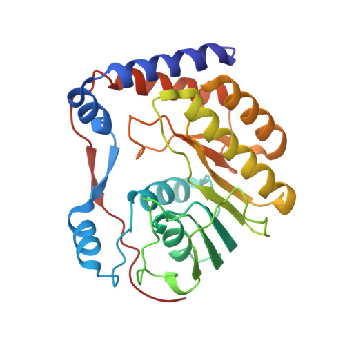
Viral methyltransferases are involved in the mRNA capping process, resulting in the transfer of a methyl group from S-adenosyl-L-methionine to capped RNA. Two groups of methyltransferases (MTases) are known: (guanine-N7)-methyltransferases (N7MTases), adding a methyl group onto the N7 atom of guanine, and (nucleoside-2'-O-)-methyltransferases (2'OMTases), adding a methyl group to a ribose hydroxyl. We have expressed and purified two constructs of Meaban virus (MV; genus Flavivirus) NS5 protein MTase domain (residues 1-265 and 1-293, respectively). We report here the three-dimensional structure of the shorter MTase construct in complex with the cofactor S-adenosyl-L-methionine, at 2.9 angstroms resolution. Inspection of the refined crystal structure, which highlights structural conservation of specific active site residues, together with sequence analysis and structural comparison with Dengue virus 2'OMTase, suggests that the crystallized enzyme belongs to the 2'OMTase subgroup. Enzymatic assays show that the short MV MTase construct is inactive, but the longer construct expressed can transfer a methyl group to the ribose 2'O atom of a short GpppAC(5) substrate. West Nile virus MTase domain has been recently shown to display both N7 and 2'O MTase activity on a capped RNA substrate comprising the 5'-terminal 190 nt of the West Nile virus genome. The lack of N7 MTase activity here reported for MV MTase may be related either to the small size of the capped RNA substrate, to its sequence, or to different structural properties of the C-terminal regions of West Nile virus and MV MTase-domains.
Organizational Affiliation:
Department of Biomolecular Sciences and Biotechnology, CNR-INFM, University of Milano, 20133-Milano, Italy.