2OXO

Crystallization and structure determination of the core-binding domain of bacteriophage lambda integrase


Experimental Data Snapshot

  • Method: X-RAY DIFFRACTION
  • Resolution: 2.00 Å
  • R-Value Free: 0.289 
  • R-Value Work: 0.228 
  • R-Value Observed: 0.234 

wwPDB Validation   3D Report Full Report


This is version 1.2 of the entry. See complete history


Literature

Crystallization and structure determination of the core-binding domain of bacteriophage lambda integrase.

Kamadurai, H.B.Jain, R.Foster, M.P.

(2008) Acta Crystallogr Sect F Struct Biol Cryst Commun 64: 470-473

  • DOI: https://doi.org/10.1107/S174430910801381X
  • Primary Citation of Related Structures:  
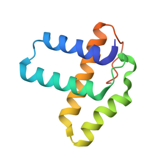
    2OXO

  • PubMed Abstract: 

    Bacteriophage lambda integrase catalyzes site-specific DNA recombination. A helical bundle domain in the enzyme, called the core-binding domain (Int(CB)), promotes the catalysis of an intermediate DNA-cleavage reaction that is critical for recombination and is not well folded in solution in the absence of DNA. To gain structural insights into the mechanism behind the accessory role of this domain in catalysis, an attempt was made to crystallize an Int(CB)-DNA complex, but crystals of free Int(CB) were fortuitously obtained. The three-dimensional structure of DNA-free Int(CB) was solved at 2.0 A resolution by molecular replacement using as the search model the previously available DNA-bound 2.8 A structure of the Int(CB) domain in a larger construct of lambda integrase. The crystal structure of DNA-free Int(CB) resembles the DNA-bound structure of Int(CB), but exhibits subtle differences in the DNA-binding face and lacks electron density for ten residues in the C-terminus that form a portion of a linker connecting Int(CB) to the C-terminal catalytic domain of the enzyme. Thus, this work reveals the domain in the absence of DNA and allows comparison with the DNA-bound form of this catalytically activating domain.


  • Organizational Affiliation

    Biophysics Program and Department of Biochemistry, The Ohio State University, Columbus, OH 43201, USA.


Macromolecules
Find similar proteins by:  (by identity cutoff)  |  3D Structure
Entity ID: 1
MoleculeChains Sequence LengthOrganismDetailsImage
Integrase103Bacteriophage sp.Mutation(s): 0 
Gene Names: int
UniProt
Find proteins for P03700 (Escherichia phage lambda)
Explore P03700 
Go to UniProtKB:  P03700
Entity Groups  
Sequence Clusters30% Identity50% Identity70% Identity90% Identity95% Identity100% Identity
UniProt GroupP03700
Sequence Annotations
Expand
  • Reference Sequence
Small Molecules
Ligands 1 Unique
IDChains Name / Formula / InChI Key2D Diagram3D Interactions
SO4
Query on SO4

Download Ideal Coordinates CCD File 
B [auth A]SULFATE ION
O4 S
QAOWNCQODCNURD-UHFFFAOYSA-L
Experimental Data & Validation

Experimental Data

  • Method: X-RAY DIFFRACTION
  • Resolution: 2.00 Å
  • R-Value Free: 0.289 
  • R-Value Work: 0.228 
  • R-Value Observed: 0.234 
  • Space Group: P 41 21 2
Unit Cell:
Length ( Å )Angle ( ˚ )
a = 46.693α = 90
b = 46.693β = 90
c = 111.533γ = 90
Software Package:
Software NamePurpose
REFMACrefinement
CrystalCleardata collection
CrystalCleardata reduction
d*TREKdata scaling
PHASERphasing

Structure Validation

View Full Validation Report


Entry History 

Deposition Data

Revision History  (Full details and data files)

  • Version 1.0: 2008-02-26
    Type: Initial release
  • Version 1.1: 2011-07-13
    Changes: Version format compliance
  • Version 1.2: 2024-02-21
    Changes: Data collection, Database references, Derived calculations