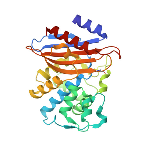
Crystal Structure of KPC-2: Insights into Carbapenemase Activity in Class A beta-Lactamases.
Ke, W., Bethel, C.R., Thomson, J.M., Bonomo, R.A., Akker, F.(2007) Biochemistry 46: 5732-5740
- PubMed: 17441734
- DOI: https://doi.org/10.1021/bi700300u
- Primary Citation of Related Structures:
2OV5 - PubMed Abstract:
Beta-lactamases inactivate beta-lactam antibiotics and are a major cause of antibiotic resistance. The recent outbreaks of Klebsiella pneumoniae carbapenem resistant (KPC) infections mediated by KPC type beta-lactamases are creating a serious threat to our "last resort" antibiotics, the carbapenems. KPC beta-lactamases are serine carbapenemases and are a subclass of class A beta-lactamases that have evolved to efficiently hydrolyze carbapenems and cephamycins which contain substitutions at the alpha-position proximal to the carbonyl group that normally render these beta-lactams resistant to hydrolysis. To investigate the molecular basis of this carbapenemase activity, we have determined the structure of KPC-2 at 1.85 A resolution. The active site of KPC-2 reveals the presence of a bicine buffer molecule which interacts via its carboxyl group with conserved active site residues S130, K234, T235, and T237; these likely resemble the interactions the beta-lactam carboxyl moiety makes in the Michaelis-Menten complex. Comparison of the KPC-2 structure with non-carbapenemases and previously determined NMC-A and SME-1 carbapenemase structures shows several active site alterations that are unique among carbapenemases. An outward shift of the catalytic S70 residue renders the active sites of the carbapenemases more shallow, likely allowing easier access of the bulkier substrates. Further space for the alpha-substituents is potentially provided by shifts in N132 and N170 in addition to concerted movements in the postulated carboxyl binding pocket that might allow the substrates to bind at a slightly different angle to accommodate these alpha-substituents. The structure of KPC-2 provides key insights into the carbapenemase activity of emerging class A beta-lactamases.
Organizational Affiliation:
Department of Biochemistry, Case Western Reserve University, Cleveland, Ohio 44106-4935, USA.