2OUZ

Crystal Structure of Estrogen Receptor alpha-lasofoxifene complex


Experimental Data Snapshot

  • Method: X-RAY DIFFRACTION
  • Resolution: 2.00 Å
  • R-Value Free: 0.269 
  • R-Value Work: 0.199 
  • R-Value Observed: 0.202 

wwPDB Validation   3D Report Full Report


Ligand Structure Quality Assessment 


This is version 1.5 of the entry. See complete history


Literature

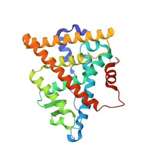
The 2.0 A crystal structure of the ER{alpha} ligand-binding domain complexed with lasofoxifene

Vajdos, F.F.Hoth, L.R.Geoghegan, K.F.Simons, S.P.LeMotte, P.K.Danley, D.E.Ammirati, M.J.Pandit, J.

(2007) Protein Sci 16: 897-905

  • DOI: https://doi.org/10.1110/ps.062729207

  • PubMed Abstract: 

    Lasofoxifene is a new and potent selective estrogen receptor modulator (SERM). The structural basis of its interaction with the estrogen receptor has been investigated by crystallographic analysis of its complex with the ligand-binding domain of estrogen receptor alpha at a resolution of 2.0 A. As with other SERMs, lasofoxifene diverts the receptor from its agonist-bound conformation by displacing the C-terminal AF-2 helix into the site at which the LXXLL motif of coactivator proteins would otherwise be able to bind. Lasofoxifene achieves this effect by occupying the space normally filled by residue Leu 540, as well as by modulating the conformation of residues of helix 11 (His 524, Leu 525). A well-defined salt bridge between lasofoxifene and Asp 351 suggests that charge neutralization in this region of the receptor may explain the some of the antiestrogenic effects of lasofoxifene. The results suggest general features of ERalpha/SERM recognition, and add a new dimension to efforts to rationalize differences between the biological activity profiles exhibited by these important pharmacological agents.


  • Organizational Affiliation

    Department of Exploratory Medicinal Sciences, Pfizer Global Research and Development, Pfizer Inc., Groton, Connecticut 06340-8001, USA. felix.vajdos@pfizer.com


Macromolecules
Find similar proteins by:  (by identity cutoff)  |  3D Structure
Entity ID: 1
MoleculeChains Sequence LengthOrganismDetailsImage
Estrogen receptor253Homo sapiensMutation(s): 0 
Gene Names: ESR1ESRNR3A1
UniProt & NIH Common Fund Data Resources
Find proteins for P03372 (Homo sapiens)
Explore P03372 
Go to UniProtKB:  P03372
PHAROS:  P03372
GTEx:  ENSG00000091831 
Entity Groups  
Sequence Clusters30% Identity50% Identity70% Identity90% Identity95% Identity100% Identity
UniProt GroupP03372
Sequence Annotations
Expand
  • Reference Sequence
Small Molecules
Ligands 1 Unique
IDChains Name / Formula / InChI Key2D Diagram3D Interactions
C3D
Query on C3D

Download Ideal Coordinates CCD File 
B [auth A](5R,6S)-6-PHENYL-5-[4-(2-PYRROLIDIN-1-YLETHOXY)PHENYL]-5,6,7,8-TETRAHYDRONAPHTHALEN-2-OL
C28 H31 N O2
GXESHMAMLJKROZ-IAPPQJPRSA-N
Binding Affinity Annotations 
IDSourceBinding Affinity
C3D BindingDB:  2OUZ IC50: min: 0.5, max: 11 (nM) from 5 assay(s)
EC50: 3.1 (nM) from 1 assay(s)
Experimental Data & Validation

Experimental Data

  • Method: X-RAY DIFFRACTION
  • Resolution: 2.00 Å
  • R-Value Free: 0.269 
  • R-Value Work: 0.199 
  • R-Value Observed: 0.202 
  • Space Group: P 65 2 2
Unit Cell:
Length ( Å )Angle ( ˚ )
a = 58.306α = 90
b = 58.306β = 90
c = 275.033γ = 120
Software Package:
Software NamePurpose
REFMACrefinement
PDB_EXTRACTdata extraction
DENZOdata reduction
SCALEPACKdata scaling
AMoREphasing

Structure Validation

View Full Validation Report


Ligand Structure Quality Assessment 


Entry History 

Deposition Data

Revision History  (Full details and data files)

  • Version 1.0: 2007-05-08
    Type: Initial release
  • Version 1.1: 2008-05-01
    Changes: Version format compliance
  • Version 1.2: 2011-07-13
    Changes: Advisory, Version format compliance
  • Version 1.3: 2018-04-04
    Changes: Data collection
  • Version 1.4: 2020-01-15
    Changes: Structure summary
  • Version 1.5: 2023-08-30
    Changes: Data collection, Database references, Derived calculations, Refinement description