2OUG

Crystal structure of the RfaH transcription factor at 2.1A resolution


Experimental Data Snapshot

  • Method: X-RAY DIFFRACTION
  • Resolution: 2.10 Å
  • R-Value Free: 0.273 
  • R-Value Work: 0.238 
  • R-Value Observed: 0.242 

wwPDB Validation   3D Report Full Report


This is version 1.4 of the entry. See complete history


Literature

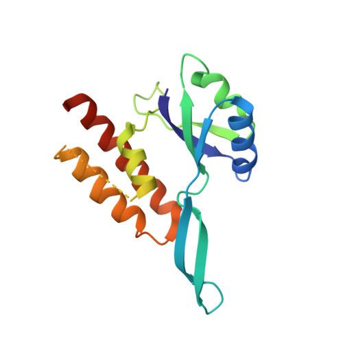
Structural basis for converting a general transcription factor into an operon-specific virulence regulator.

Belogurov, G.A.Vassylyeva, M.N.Svetlov, V.Klyuyev, S.Grishin, N.V.Vassylyev, D.G.Artsimovitch, I.

(2007) Mol Cell 26: 117-129

  • DOI: https://doi.org/10.1016/j.molcel.2007.02.021
  • Primary Citation of Related Structures:  
    2OUG

  • PubMed Abstract: 

    RfaH, a paralog of the general transcription factor NusG, is recruited to elongating RNA polymerase at specific regulatory sites. The X-ray structure of Escherichia coli RfaH reported here reveals two domains. The N-terminal domain displays high similarity to that of NusG. In contrast, the alpha-helical coiled-coil C domain, while retaining sequence similarity, is strikingly different from the beta barrel of NusG. To our knowledge, such an all-beta to all-alpha transition of the entire domain is the most extreme example of protein fold evolution known to date. Both N domains possess a vast hydrophobic cavity that is buried by the C domain in RfaH but is exposed in NusG. We propose that this cavity constitutes the RNA polymerase-binding site, which becomes unmasked in RfaH only upon sequence-specific binding to the nontemplate DNA strand that triggers domain dissociation. Finally, we argue that RfaH binds to the beta' subunit coiled coil, the major target site for the initiation sigma factors.


  • Organizational Affiliation

    Department of Microbiology, The Ohio State University, Columbus, OH 43210, USA.


Macromolecules
Find similar proteins by:  (by identity cutoff)  |  3D Structure
Entity ID: 1
MoleculeChains Sequence LengthOrganismDetailsImage
Transcriptional activator rfaH
A, B, C, D
162Escherichia coliMutation(s): 0 
Gene Names: rfaHhlyTsfrB
UniProt
Find proteins for P0AFW0 (Escherichia coli (strain K12))
Explore P0AFW0 
Go to UniProtKB:  P0AFW0
Entity Groups  
Sequence Clusters30% Identity50% Identity70% Identity90% Identity95% Identity100% Identity
UniProt GroupP0AFW0
Sequence Annotations
Expand
  • Reference Sequence
Experimental Data & Validation

Experimental Data

  • Method: X-RAY DIFFRACTION
  • Resolution: 2.10 Å
  • R-Value Free: 0.273 
  • R-Value Work: 0.238 
  • R-Value Observed: 0.242 
  • Space Group: P 65
Unit Cell:
Length ( Å )Angle ( ˚ )
a = 45.149α = 90
b = 45.149β = 90
c = 600.163γ = 120
Software Package:
Software NamePurpose
MAR345data collection
MLPHAREphasing
CNSrefinement
HKL-2000data reduction
HKL-2000data scaling

Structure Validation

View Full Validation Report


Entry History 

Deposition Data

Revision History  (Full details and data files)

  • Version 1.0: 2007-05-01
    Type: Initial release
  • Version 1.1: 2008-05-01
    Changes: Version format compliance
  • Version 1.2: 2011-07-13
    Changes: Version format compliance
  • Version 1.3: 2017-10-18
    Changes: Refinement description
  • Version 1.4: 2024-02-21
    Changes: Data collection, Database references