2OUA

Crystal Structure of Nocardiopsis Protease (NAPase)


Experimental Data Snapshot

  • Method: X-RAY DIFFRACTION
  • Resolution: 1.85 Å
  • R-Value Free: 0.200 
  • R-Value Work: 0.176 

wwPDB Validation   3D Report Full Report


Ligand Structure Quality Assessment 


This is version 1.4 of the entry. See complete history


Literature

Structural and mechanistic exploration of Acid resistance: kinetic stability facilitates evolution of extremophilic behavior

Kelch, B.A.Eagen, K.P.Erciyas, F.P.Humphris, E.L.Thomason, A.R.Mitsuiki, S.Agard, D.A.

(2007) J Mol Biol 368: 870-883

  • DOI: https://doi.org/10.1016/j.jmb.2007.02.032
  • Primary Citation of Related Structures:  
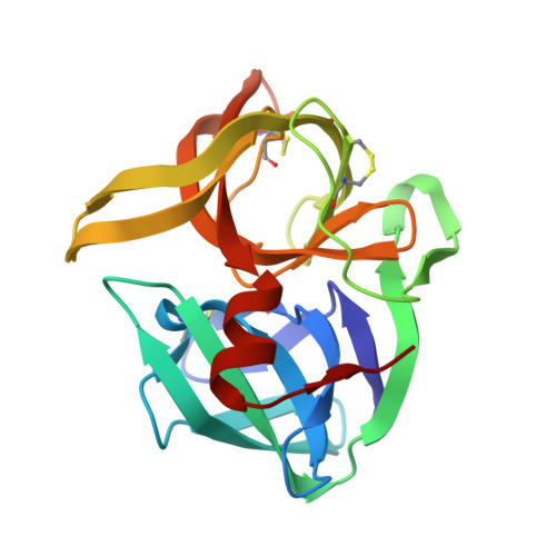
    2OUA

  • PubMed Abstract: 

    Kinetically stable proteins are unique in that their stability is determined solely by kinetic barriers rather than by thermodynamic equilibria. To better understand how kinetic stability promotes protein survival under extreme environmental conditions, we analyzed the unfolding behavior and determined the structure of Nocardiopsis alba Protease A (NAPase), an acid-resistant, kinetically stable protease, and compared these results with a neutrophilic homolog, alpha-lytic protease (alphaLP). Although NAPase and alphaLP have the same number of acid-titratable residues, kinetic studies revealed that the height of the unfolding free energy barrier for NAPase is less sensitive to acid than that of alphaLP, thereby accounting for NAPase's improved tolerance of low pH. A comparison of the alphaLP and NAPase structures identified multiple salt-bridges in the domain interface of alphaLP that were relocated to outer regions of NAPase, suggesting a novel mechanism of acid stability in which acid-sensitive electrostatic interactions are rearranged to similarly affect the energetics of both the native state and the unfolding transition state. An acid-stable variant of alphaLP in which a single interdomain salt-bridge is replaced with a corresponding intradomain NAPase salt-bridge shows a dramatic >15-fold increase in acid resistance, providing further evidence for this hypothesis. These observations also led to a general model of the unfolding transition state structure for alphaLP protease family members in which the two domains separate from each other while remaining relatively intact themselves. These results illustrate the remarkable utility of kinetic stability as an evolutionary tool for developing longevity over a broad range of harsh conditions.


  • Organizational Affiliation

    Howard Hughes Medical Institute and the Department of Biochemistry and Biophysics, University of California-San Francisco, 600 16th Street, San Francisco, CA 94158-2517, USA.


Macromolecules
Find similar proteins by:  (by identity cutoff)  |  3D Structure
Entity ID: 1
MoleculeChains Sequence LengthOrganismDetailsImage
Serine protease
A, B
188Nocardiopsis albaMutation(s): 0 
UniProt
Find proteins for Q6K1C5 (Nocardiopsis sp. TOA-1)
Explore Q6K1C5 
Go to UniProtKB:  Q6K1C5
Entity Groups  
Sequence Clusters30% Identity50% Identity70% Identity90% Identity95% Identity100% Identity
UniProt GroupQ6K1C5
Sequence Annotations
Expand
  • Reference Sequence
Small Molecules
Ligands 4 Unique
IDChains Name / Formula / InChI Key2D Diagram3D Interactions
AES
Query on AES

Download Ideal Coordinates CCD File 
E [auth A],
J [auth B]
4-(2-AMINOETHYL)BENZENESULFONYL FLUORIDE
C8 H10 F N O2 S
MGSKVZWGBWPBTF-UHFFFAOYSA-N
SO4
Query on SO4

Download Ideal Coordinates CCD File 
C [auth A],
D [auth A]
SULFATE ION
O4 S
QAOWNCQODCNURD-UHFFFAOYSA-L
GOL
Query on GOL

Download Ideal Coordinates CCD File 
H [auth A],
I [auth A],
L [auth B]
GLYCEROL
C3 H8 O3
PEDCQBHIVMGVHV-UHFFFAOYSA-N
DIO
Query on DIO

Download Ideal Coordinates CCD File 
F [auth A],
G [auth A],
K [auth B]
1,4-DIETHYLENE DIOXIDE
C4 H8 O2
RYHBNJHYFVUHQT-UHFFFAOYSA-N
Experimental Data & Validation

Experimental Data

  • Method: X-RAY DIFFRACTION
  • Resolution: 1.85 Å
  • R-Value Free: 0.200 
  • R-Value Work: 0.176 
  • Space Group: P 32 2 1
Unit Cell:
Length ( Å )Angle ( ˚ )
a = 61.605α = 90
b = 61.605β = 90
c = 184.479γ = 120
Software Package:
Software NamePurpose
HKL-2000data collection
CNSrefinement
DENZOdata reduction
SCALEPACKdata scaling
CNSphasing

Structure Validation

View Full Validation Report


Ligand Structure Quality Assessment 


Entry History 

Deposition Data

Revision History  (Full details and data files)

  • Version 1.0: 2007-02-20
    Type: Initial release
  • Version 1.1: 2008-05-01
    Changes: Version format compliance
  • Version 1.2: 2011-07-13
    Changes: Non-polymer description, Version format compliance
  • Version 1.3: 2012-06-06
    Changes: Non-polymer description
  • Version 1.4: 2024-04-03
    Changes: Data collection, Database references, Derived calculations, Refinement description