


Crystal structure and inhibition of a catalytically active form of diaminopimelate epimerase (DapF)from Bacillus anthracis
Matho, M.H., Fukuda, K., Lloyd, A.J., Santelli, E., Jaroszewski, L., Scott, D.J., Liddington, R.C., Roper, D.To be published.
Experimental Data Snapshot
wwPDB Validation 3D Report Full Report

Entity ID: 1 | |||||
|---|---|---|---|---|---|
| Molecule | Chains | Sequence Length | Organism | Details | Image |
| Diaminopimelate epimerase | 308 | Bacillus anthracis str. Ames | Mutation(s): 0 Gene Names: dapF EC: 5.1.1.7 | 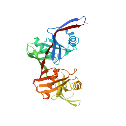 | |
UniProt | |||||
Find proteins for Q81XR2 (Bacillus anthracis) Explore Q81XR2 Go to UniProtKB: Q81XR2 | |||||
Entity Groups | |||||
| Sequence Clusters | 30% Identity50% Identity70% Identity90% Identity95% Identity100% Identity | ||||
| UniProt Group | Q81XR2 | ||||
Sequence AnnotationsExpand | |||||
| |||||
| Length ( Å ) | Angle ( ˚ ) |
|---|---|
| a = 64.863 | α = 90 |
| b = 87.334 | β = 90 |
| c = 110.453 | γ = 90 |
| Software Name | Purpose |
|---|---|
| REFMAC | refinement |
| DENZO | data reduction |
| SCALEPACK | data scaling |
| MOLREP | phasing |
RCSB PDB (citation) is hosted by
RCSB PDB is a member of the

