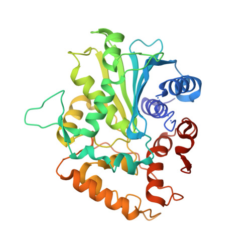
Structural basis for the cold adaptation of psychrophilic M37 lipase from Photobacterium lipolyticum.
Jung, S.K., Jeong, D.G., Lee, M.S., Lee, J.K., Kim, H.K., Ryu, S.E., Park, B.C., Kim, J.H., Kim, S.J.(2008) Proteins 71: 476-484
- PubMed: 18186467
- DOI: https://doi.org/10.1002/prot.21884
- Primary Citation of Related Structures:
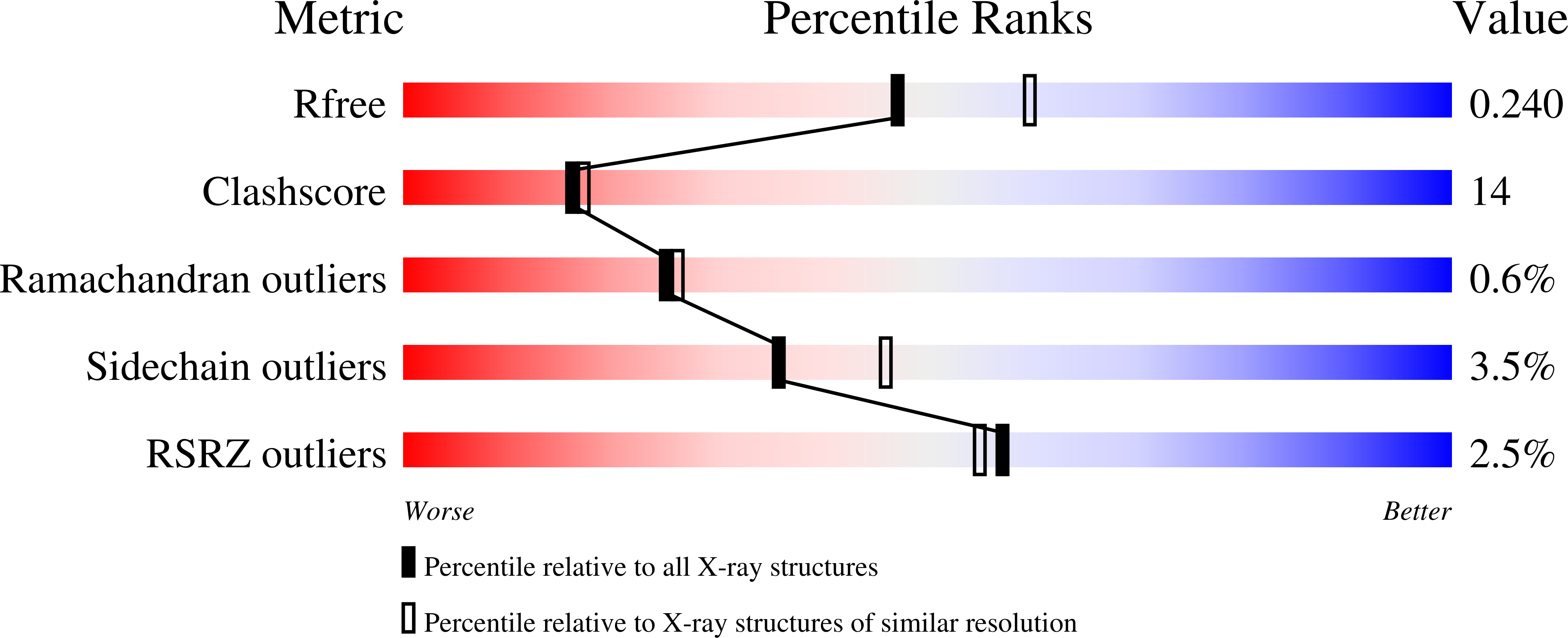
2ORY - PubMed Abstract:
The M37 lipase from Photobacterium lipolyticum shows an extremely low activation energy and strong activity at low temperatures, with optimum activity seen at 298 K and more than 75% of the optimum activity retained down to 278 K. Though the M37 lipase is most closely related to the filamentous fungal lipase, Rhizomucor miehei lipase (RML) at the primary structure level, their activity characteristics are completely different. In an effort to identify structural components of cold adaptation in lipases, we determined the crystal structure of the M37 lipase at 2.2 A resolution and compared it to that of nonadapted RML. Structural analysis revealed that M37 lipase adopted a folding pattern similar to that observed for other lipase structures. However, comparison with RML revealed that the region beneath the lid of the M37 lipase included a significant and unique cavity that would be occupied by a lid helix upon substrate binding. In addition, the oxyanion hole was much wider in M37 lipase than RML. We propose that these distinct structural characteristics of M37 lipase may facilitate the lateral movement of the helical lid and subsequent substrate hydrolysis, which might explain its low activation energy and high activity at low temperatures.
Organizational Affiliation:
Translational Research Center, Korea Research Institute of Bioscience and Biotechnology, Yuseong-Gu, Daejeon 305-600, Korea.