


2OR4
A high resolution crystal structure of human glutamate carboxypeptidase II in complex with quisqualic acid
- PDB DOI: https://doi.org/10.2210/pdb2OR4/pdb
- Classification: HYDROLASE
- Organism(s): Homo sapiens
- Expression System: Drosophila melanogaster
- Mutation(s): No
- Deposited: 2007-02-01 Released: 2007-06-12
Experimental Data Snapshot
- Method: X-RAY DIFFRACTION
- Resolution: 1.62 Å
- R-Value Free: 0.219
- R-Value Work: 0.184
- R-Value Observed: 0.186

This is version 2.1 of the entry. See complete history.

Macromolecules
Find similar proteins by:
(by identity cutoff) | 3D Structure
Entity ID: 1 | |||||
|---|---|---|---|---|---|
| Molecule | Chains | Sequence Length | Organism | Details | Image |
| Glutamate carboxypeptidase 2 | 709 | Homo sapiens | Mutation(s): 0 Gene Names: FOLH1, FOLH, NAALAD1, PSM, PSMA EC: 3.4.17.21 | 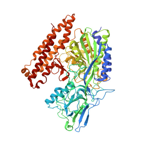 | |
UniProt & NIH Common Fund Data Resources | |||||
Find proteins for Q04609 (Homo sapiens) Explore Q04609 Go to UniProtKB: Q04609 | |||||
PHAROS: Q04609 GTEx: ENSG00000086205 | |||||
Entity Groups | |||||
| Sequence Clusters | 30% Identity50% Identity70% Identity90% Identity95% Identity100% Identity | ||||
| UniProt Group | Q04609 | ||||
Sequence AnnotationsExpand | |||||
| |||||
Oligosaccharides
Entity ID: 2 | |||||
|---|---|---|---|---|---|
| Molecule | Chains | Length | 2D Diagram | Glycosylation | 3D Interactions |
| 2-acetamido-2-deoxy-beta-D-glucopyranose-(1-4)-2-acetamido-2-deoxy-beta-D-glucopyranose | B, C, D | 2 |  | N-Glycosylation | |
Glycosylation Resources | |||||
GlyTouCan: G42666HT GlyCosmos: G42666HT GlyGen: G42666HT | |||||
Entity ID: 3 | |||||
|---|---|---|---|---|---|
| Molecule | Chains | Length | 2D Diagram | Glycosylation | 3D Interactions |
| alpha-D-mannopyranose-(1-3)-beta-D-mannopyranose-(1-4)-2-acetamido-2-deoxy-beta-D-glucopyranose-(1-4)-2-acetamido-2-deoxy-beta-D-glucopyranose | E | 4 |  | N-Glycosylation | |
Glycosylation Resources | |||||
GlyTouCan: G81315DD GlyCosmos: G81315DD GlyGen: G81315DD | |||||
Small Molecules
| Ligands 5 Unique | |||||
|---|---|---|---|---|---|
| ID | Chains | Name / Formula / InChI Key | 2D Diagram | 3D Interactions | |
| NAG Query on NAG | F [auth A], G [auth A], H [auth A] | 2-acetamido-2-deoxy-beta-D-glucopyranose C8 H15 N O6 OVRNDRQMDRJTHS-FMDGEEDCSA-N |  | ||
| QUS Query on QUS | M [auth A] | (S)-2-AMINO-3-(3,5-DIOXO-[1,2,4]OXADIAZOLIDIN-2-YL)-PROPIONIC ACID C5 H7 N3 O5 ASNFTDCKZKHJSW-REOHCLBHSA-N |  | ||
| ZN Query on ZN | I [auth A], J [auth A] | ZINC ION Zn PTFCDOFLOPIGGS-UHFFFAOYSA-N |  | ||
| CA Query on CA | K [auth A] | CALCIUM ION Ca BHPQYMZQTOCNFJ-UHFFFAOYSA-N |  | ||
| CL Query on CL | L [auth A] | CHLORIDE ION Cl VEXZGXHMUGYJMC-UHFFFAOYSA-M |  | ||
Experimental Data & Validation
Experimental Data
- Method: X-RAY DIFFRACTION
- Resolution: 1.62 Å
- R-Value Free: 0.219
- R-Value Work: 0.184
- R-Value Observed: 0.186
- Space Group: I 2 2 2
Unit Cell:
| Length ( Å ) | Angle ( ˚ ) |
|---|---|
| a = 102.007 | α = 90 |
| b = 130.437 | β = 90 |
| c = 159.508 | γ = 90 |
| Software Name | Purpose |
|---|---|
| REFMAC | refinement |
| MAR345dtb | data collection |
| HKL-2000 | data reduction |
| HKL-2000 | data scaling |
Entry History
Deposition Data
- Released Date: 2007-06-12 Deposition Author(s): Barinka, C., Lubkowski, J.
Revision History (Full details and data files)
- Version 1.0: 2007-06-12
Type: Initial release - Version 1.1: 2007-10-24
Changes: Version format compliance - Version 1.2: 2011-07-13
Changes: Derived calculations, Version format compliance - Version 2.0: 2020-07-29
Type: Remediation
Reason: Carbohydrate remediation
Changes: Advisory, Atomic model, Data collection, Database references, Derived calculations, Structure summary - Version 2.1: 2023-08-30
Changes: Data collection, Database references, Refinement description, Structure summary
