2OQX

Crystal Structure of the apo form of E. coli tryptophanase at 1.9 A resolution


Experimental Data Snapshot

  • Method: X-RAY DIFFRACTION
  • Resolution: 1.90 Å
  • R-Value Free: 0.232 
  • R-Value Work: 0.203 
  • R-Value Observed: 0.210 

wwPDB Validation   3D Report Full Report


This is version 1.5 of the entry. See complete history


Literature

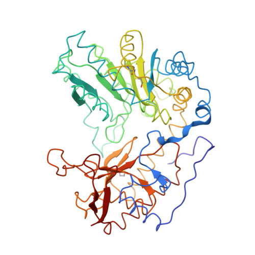
The structure of apo tryptophanase from Escherichia coli reveals a wide-open conformation.

Tsesin, N.Kogan, A.Gdalevsky, G.Y.Himanen, J.P.Cohen-Luria, R.Parola, A.H.Goldgur, Y.Almog, O.

(2007) Acta Crystallogr D Biol Crystallogr 63: 969-974

  • DOI: https://doi.org/10.1107/S0907444907036396
  • Primary Citation of Related Structures:  
    2OQX

  • PubMed Abstract: 

    The crystal structure of apo tryptophanase from Escherichia coli (space group F222, unit-cell parameters a = 118.4, b = 120.1, c = 171.2 A) was determined at 1.9 A resolution using the molecular-replacement method and refined to an R factor of 20.3% (R(free) = 23.2%). The structure revealed a significant shift in the relative orientation of the domains compared with both the holo form of Proteus vulgaris tryptophanase and with another crystal structure of apo E. coli tryptophanase, reflecting the internal flexibility of the molecule. Domain shifts were previously observed in tryptophanase and in the closely related enzyme tyrosine phenol-lyase, with the holo form found in an open conformation and the apo form in either an open or a closed conformation. Here, a wide-open conformation of the apo form of tryptophanase is reported. A conformational change is also observed in loop 297-303. The structure contains a hydrated Mg(2+) at the cation-binding site and a Cl(-) ion at the subunit interface. The enzyme activity depends on the nature of the bound cation, with smaller ions serving as inhibitors. It is hypothesized that this effect arises from variations of the coordination geometry of the bound cation.


  • Organizational Affiliation

    Department of Chemistry, Ben-Gurion University of the Negev, POB 653, Beer-Sheva 84105, Israel.


Macromolecules
Find similar proteins by:  (by identity cutoff)  |  3D Structure
Entity ID: 1
MoleculeChains Sequence LengthOrganismDetailsImage
Tryptophanase467Escherichia coliMutation(s): 2 
EC: 4.1.99.1
UniProt
Find proteins for Q5UES8 (Escherichia coli)
Explore Q5UES8 
Go to UniProtKB:  Q5UES8
Entity Groups  
Sequence Clusters30% Identity50% Identity70% Identity90% Identity95% Identity100% Identity
UniProt GroupQ5UES8
Sequence Annotations
Expand
  • Reference Sequence
Small Molecules
Modified Residues  1 Unique
IDChains TypeFormula2D DiagramParent
CME
Query on CME
A
L-PEPTIDE LINKINGC5 H11 N O3 S2CYS
Experimental Data & Validation

Experimental Data

  • Method: X-RAY DIFFRACTION
  • Resolution: 1.90 Å
  • R-Value Free: 0.232 
  • R-Value Work: 0.203 
  • R-Value Observed: 0.210 
  • Space Group: F 2 2 2
Unit Cell:
Length ( Å )Angle ( ˚ )
a = 118.422α = 90
b = 120.116β = 90
c = 171.209γ = 90
Software Package:
Software NamePurpose
CNSrefinement
PDB_EXTRACTdata extraction
MAR345data collection
HKL-2000data reduction
SCALEPACKdata scaling
REFMACrefinement

Structure Validation

View Full Validation Report


Entry History 

Deposition Data

Revision History  (Full details and data files)

  • Version 1.0: 2007-02-20
    Type: Initial release
  • Version 1.1: 2007-10-24
    Changes: Version format compliance
  • Version 1.2: 2011-07-13
    Changes: Derived calculations, Version format compliance
  • Version 1.3: 2017-10-18
    Changes: Refinement description
  • Version 1.4: 2019-07-24
    Changes: Data collection, Derived calculations, Refinement description
  • Version 1.5: 2023-12-27
    Changes: Data collection, Database references, Derived calculations