2OP3

The structure of cathepsin S with a novel 2-arylphenoxyacetaldehyde inhibitor derived by the Substrate Activity Screening (SAS) method


Experimental Data Snapshot

  • Method: X-RAY DIFFRACTION
  • Resolution: 1.60 Å
  • R-Value Free: 0.181 
  • R-Value Work: 0.158 
  • R-Value Observed: 0.159 

wwPDB Validation   3D Report Full Report


This is version 1.3 of the entry. See complete history


Literature

Characterization and optimization of selective, nonpeptidic inhibitors of cathepsin S with an unprecedented binding mode.

Inagaki, H.Tsuruoka, H.Hornsby, M.Lesley, S.A.Spraggon, G.Ellman, J.A.

(2007) J Med Chem 50: 2693-2699

  • DOI: https://doi.org/10.1021/jm070111+
  • Primary Citation of Related Structures:  
    2OP3

  • PubMed Abstract: 

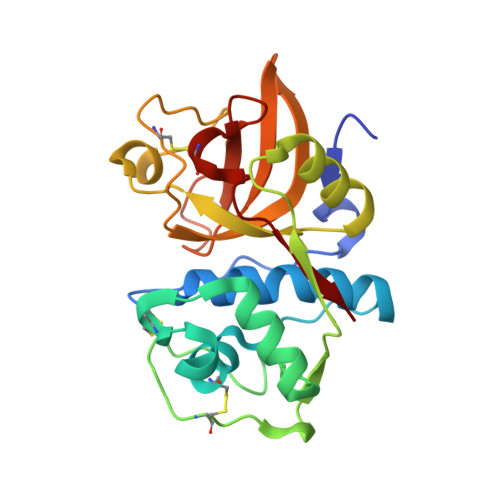
    The substrate activity screening (SAS) method, a substrate-based fragment identification and optimization method for the development of enzyme inhibitors, was previously applied to cathepsin S to obtain a novel (2-arylphenoxy)acetaldehyde inhibitor, 2, with a 0.49 microM Ki value (Wood, W. J. L.; Patterson, A. W.; Tsuruoka, H.; Jain, R. K.; Ellman, J. A. J. Am. Chem. Soc. 2005, 127, 15521-15527). In this paper we disclose the X-ray structure of a complex between cathepsin S and inhibitor 2 which reveals an unprecedented binding mode. On the basis of this structure, additional 2-biaryloxy substrates with greatly increased cleavage efficiency were designed. Conversion of the optimized substrates to the corresponding aldehyde inhibitors yielded a low molecular weight (304 Daltons) and potent (9.6 nM) cathepsin S inhibitor that showed from 100- to >1000-fold selectivity relative to cathepsins B, L, and K.


  • Organizational Affiliation

    Department of Chemistry, University of California, Berkeley, California 94720, USA.


Macromolecules
Find similar proteins by:  (by identity cutoff)  |  3D Structure
Entity ID: 1
MoleculeChains Sequence LengthOrganismDetailsImage
Cathepsin S
A, B
220Homo sapiensMutation(s): 0 
Gene Names: CATHS
EC: 3.4.22.27
UniProt & NIH Common Fund Data Resources
Find proteins for P25774 (Homo sapiens)
Explore P25774 
Go to UniProtKB:  P25774
PHAROS:  P25774
GTEx:  ENSG00000163131 
Entity Groups  
Sequence Clusters30% Identity50% Identity70% Identity90% Identity95% Identity100% Identity
UniProt GroupP25774
Sequence Annotations
Expand
  • Reference Sequence
Small Molecules
Ligands 3 Unique
IDChains Name / Formula / InChI Key2D Diagram3D Interactions
PEU
Query on PEU

Download Ideal Coordinates CCD File 
E [auth A]2,5,8,11,14,17,20,23,26,29,32,35,38,41,44,47,50,53,56,59,62,65,68,71,74,77,80-HEPTACOSAOXADOOCTACONTAN-82-OL
C55 H112 O28
ISGUIIHZEJGUGQ-UHFFFAOYSA-N
TF5
Query on TF5

Download Ideal Coordinates CCD File 
D [auth A],
F [auth B]
2-[(2',3',4'-TRIFLUOROBIPHENYL-2-YL)OXY]ETHANOL
C14 H11 F3 O2
AGXPXFPIOHNDAN-UHFFFAOYSA-N
SO4
Query on SO4

Download Ideal Coordinates CCD File 
C [auth A]SULFATE ION
O4 S
QAOWNCQODCNURD-UHFFFAOYSA-L
Binding Affinity Annotations 
IDSourceBinding Affinity
TF5 PDBBind:  2OP3 Ki: 490 (nM) from 1 assay(s)
Experimental Data & Validation

Experimental Data

  • Method: X-RAY DIFFRACTION
  • Resolution: 1.60 Å
  • R-Value Free: 0.181 
  • R-Value Work: 0.158 
  • R-Value Observed: 0.159 
  • Space Group: P 41 2 2
Unit Cell:
Length ( Å )Angle ( ˚ )
a = 85.384α = 90
b = 85.384β = 90
c = 151.2γ = 90
Software Package:
Software NamePurpose
REFMACrefinement
HKL-2000data collection
HKL-2000data reduction
HKL-2000data scaling
FFTphasing

Structure Validation

View Full Validation Report


Entry History 

Deposition Data

Revision History  (Full details and data files)

  • Version 1.0: 2007-05-22
    Type: Initial release
  • Version 1.1: 2008-05-01
    Changes: Version format compliance
  • Version 1.2: 2011-07-13
    Changes: Version format compliance
  • Version 1.3: 2023-08-30
    Changes: Data collection, Database references, Derived calculations, Refinement description