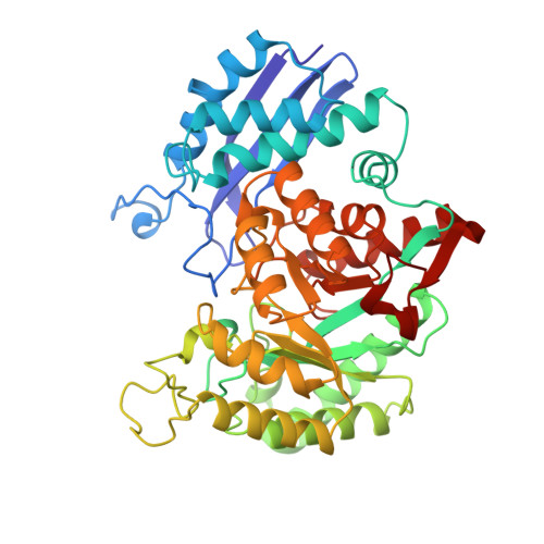
Mechanism of enolase: the crystal structure of asymmetric dimer enolase-2-phospho-D-glycerate/enolase-phosphoenolpyruvate at 2.0 A resolution.
Zhang, E., Brewer, J.M., Minor, W., Carreira, L.A., Lebioda, L.(1997) Biochemistry 36: 12526-12534
- PubMed: 9376357
- DOI: https://doi.org/10.1021/bi9712450
- Primary Citation of Related Structures:
2ONE - PubMed Abstract:
Enolase, a glycolytic enzyme that catalyzes the dehydration of 2-phospho-d-glycerate (PGA) to form phosphoenolpyruvate (PEP), is a homodimer in all eukaryotes and many prokaryotes. Here, we report the crystal structure of a complex between yeast enolase and an equilibrium mixture of PGA and PEP. The structure has been refined using 29 854 reflections with an F/sigma(F) of >/=3 to an R of 0.137 with average deviations of bond lengths and bond angles from ideal values of 0.013 A and 3.1 degrees , respectively. In this structure, the dimer constitutes the crystallographic asymmetric unit. The two subunits are similar, and their superposition gives a rms distance between Calpha atoms of 0.91 A. The exceptions to this are the catalytic loop Val153-Phe169 where the atomic positions in the two subunits differ by up to 4 A and the loop Ser250-Gln277, which follows the catalytic loop Val153-Phe169. In the first subunit, the imidazole side chain of His159 is in contact with the phosphate group of the substrate/product molecule; in the other it is separated by water molecules. A series of hydrogen bonds leading to a neighboring enolase dimer can be identified as being responsible for ordering and stabilization of the conformationally different subunits in the crystal lattice. The electron density present in the active site suggests that in the active site with the direct ligand-His159 hydrogen bond PGA is predominantly bound while in the active site where water molecules separate His159 from the ligand the binding of PEP dominates. The structure indicates that the water molecule hydrating carbon-3 of PEP in the PEP --> PGA reaction is activated by the carboxylates of Glu168 and Glu211. The crystals are unique because they have resolved two intermediates on the opposite sides of the transition state.
Organizational Affiliation:
Department of Chemistry and Biochemistry, University of South Carolina, Columbia, South Carolina 29208, USA.