2OLZ

Structure of human insulin in presence of thiocyanate at pH 7.0


Experimental Data Snapshot

  • Method: X-RAY DIFFRACTION
  • Resolution: 1.70 Å
  • R-Value Free: 0.221 
  • R-Value Work: 0.178 
  • R-Value Observed: 0.181 

wwPDB Validation   3D Report Full Report


This is version 1.4 of the entry. See complete history


Literature

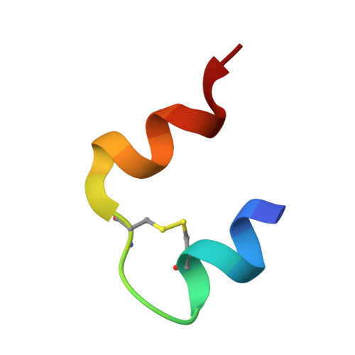
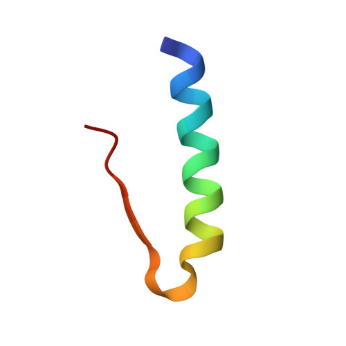
Crystallographic characterization of two novel crystal forms of human insulin induced by chaotropic agents and a shift in pH.

Norrman, M.Schluckebier, G.

(2007) BMC Struct Biol 7: 83-83

  • DOI: https://doi.org/10.1186/1472-6807-7-83
  • Primary Citation of Related Structures:  
    2OLY, 2OLZ, 2OM0, 2OM1

  • PubMed Abstract: 

    Insulin is a therapeutic protein that is widely used for the treatment of diabetes. Its biological function was discovered more than 80 years ago and it has since then been characterized extensively. Crystallization of the insulin molecule has always been a key activity since the protein is often administered by subcutaneous injections of crystalline insulin formulations. Over the years, insulin has been crystallized and characterized in a number of crystal systems.


  • Organizational Affiliation

    Diabetes Protein Engineering, Novo Nordisk A/S, Novo Nordisk Park, DK-2760 Måløv, Denmark. mtno@novonordisk.com


Macromolecules

Find similar proteins by:  Sequence   |   3D Structure  

Entity ID: 1
MoleculeChains Sequence LengthOrganismDetailsImage
Insulin A
A, C, E, G, I
A, C, E, G, I, K
21Homo sapiensMutation(s): 0 
UniProt & NIH Common Fund Data Resources
Find proteins for P01308 (Homo sapiens)
Explore P01308 
Go to UniProtKB:  P01308
PHAROS:  P01308
GTEx:  ENSG00000254647 
Entity Groups  
Sequence Clusters30% Identity50% Identity70% Identity90% Identity95% Identity100% Identity
UniProt GroupP01308
Sequence Annotations
Expand
  • Reference Sequence
Find similar proteins by:  (by identity cutoff)  |  3D Structure
Entity ID: 2
MoleculeChains Sequence LengthOrganismDetailsImage
Insulin B
B, D, F, H, J
B, D, F, H, J, L
30Homo sapiensMutation(s): 0 
UniProt & NIH Common Fund Data Resources
Find proteins for P01308 (Homo sapiens)
Explore P01308 
Go to UniProtKB:  P01308
PHAROS:  P01308
GTEx:  ENSG00000254647 
Entity Groups  
Sequence Clusters30% Identity50% Identity70% Identity90% Identity95% Identity100% Identity
UniProt GroupP01308
Sequence Annotations
Expand
  • Reference Sequence
Small Molecules
Ligands 4 Unique
IDChains Name / Formula / InChI Key2D Diagram3D Interactions
RCO
Query on RCO

Download Ideal Coordinates CCD File 
M [auth A]
P [auth C]
Q [auth E]
R [auth G]
U [auth I]
M [auth A],
P [auth C],
Q [auth E],
R [auth G],
U [auth I],
V [auth K]
RESORCINOL
C6 H6 O2
GHMLBKRAJCXXBS-UHFFFAOYSA-N
GOL
Query on GOL

Download Ideal Coordinates CCD File 
W [auth K]GLYCEROL
C3 H8 O3
PEDCQBHIVMGVHV-UHFFFAOYSA-N
ZN
Query on ZN

Download Ideal Coordinates CCD File 
N [auth B],
S [auth H]
ZINC ION
Zn
PTFCDOFLOPIGGS-UHFFFAOYSA-N
SCN
Query on SCN

Download Ideal Coordinates CCD File 
O [auth B],
T [auth H]
THIOCYANATE ION
C N S
ZMZDMBWJUHKJPS-UHFFFAOYSA-M
Experimental Data & Validation

Experimental Data

  • Method: X-RAY DIFFRACTION
  • Resolution: 1.70 Å
  • R-Value Free: 0.221 
  • R-Value Work: 0.178 
  • R-Value Observed: 0.181 
  • Space Group: C 1 2 1
Unit Cell:
Length ( Å )Angle ( ˚ )
a = 100.6α = 90
b = 60.8β = 116.1
c = 62.1γ = 90
Software Package:
Software NamePurpose
REFMACrefinement
XDSdata reduction
XSCALEdata scaling
MOLREPphasing

Structure Validation

View Full Validation Report


Entry History 

Deposition Data

Revision History  (Full details and data files)

  • Version 1.0: 2007-12-04
    Type: Initial release
  • Version 1.1: 2011-07-13
    Changes: Advisory, Derived calculations, Version format compliance
  • Version 1.2: 2018-03-07
    Changes: Data collection
  • Version 1.3: 2023-12-27
    Changes: Data collection, Database references, Derived calculations
  • Version 1.4: 2024-04-03
    Changes: Refinement description