2OKL

Crystal structure of Peptide Deformylase 2 with actinonin from Bacillus cereus


Experimental Data Snapshot

  • Method: X-RAY DIFFRACTION
  • Resolution: 1.70 Å
  • R-Value Free: 0.257 
  • R-Value Work: 0.228 
  • R-Value Observed: 0.228 

wwPDB Validation   3D Report Full Report


This is version 1.2 of the entry. See complete history


Literature

Characterization of Peptide Deformylase2 from B. cereus

Park, J.K.Kim, K.H.Moon, J.H.Kim, E.E.

(2007) J Biochem Mol Biol 40: 1050-1057

  • DOI: https://doi.org/10.5483/bmbrep.2007.40.6.1050
  • Primary Citation of Related Structures:  
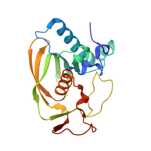
    2OKL

  • PubMed Abstract: 

    Peptide deformylase (PDF) is a metalloenzyme that removes the N-terminal formyl groups from newly synthesized proteins. It is essential for bacterial survival, and is therefore-considered as a potential target for antimicrobial chemotherapy. However, some bacteria including medically relevant pathogens possess two or more def-like genes. Here we have examined two PDFs from Bacillus cereus. The two share only 32% sequence identity and the crystal structures show overall similarity with PDF2 having a longer C-terminus. However, there are differences at the two active sites, and these differences appear to contribute to the activity difference seen between the two. BcPDF2 is found as a dimer in the crystal form with two additional actinonin bound at that interface.


  • Organizational Affiliation

    Life Sciences Division, Korea Institute of Science and Technology, 39-1 Hawolkok-dong, Sungbuk-gu, Seoul 136-791, Korea.


Macromolecules
Find similar proteins by:  (by identity cutoff)  |  3D Structure
Entity ID: 1
MoleculeChains Sequence LengthOrganismDetailsImage
Peptide deformylase 2
A, B
185Bacillus cereus ATCC 14579Mutation(s): 0 
Gene Names: def2
EC: 3.5.1.88
UniProt
Find proteins for Q819K2 (Bacillus cereus (strain ATCC 14579 / DSM 31 / CCUG 7414 / JCM 2152 / NBRC 15305 / NCIMB 9373 / NCTC 2599 / NRRL B-3711))
Explore Q819K2 
Go to UniProtKB:  Q819K2
Entity Groups  
Sequence Clusters30% Identity50% Identity70% Identity90% Identity95% Identity100% Identity
UniProt GroupQ819K2
Sequence Annotations
Expand
  • Reference Sequence
Experimental Data & Validation

Experimental Data

  • Method: X-RAY DIFFRACTION
  • Resolution: 1.70 Å
  • R-Value Free: 0.257 
  • R-Value Work: 0.228 
  • R-Value Observed: 0.228 
  • Space Group: P 65 2 2
Unit Cell:
Length ( Å )Angle ( ˚ )
a = 69.447α = 90
b = 69.447β = 90
c = 294.412γ = 120
Software Package:
Software NamePurpose
CNSrefinement
HKL-2000data collection
HKL-2000data reduction
HKL-2000data scaling
CNSphasing

Structure Validation

View Full Validation Report


Entry History 

Deposition Data

  • Released Date: 2008-01-15 
  • Deposition Author(s): Kim, E.E.

Revision History  (Full details and data files)

  • Version 1.0: 2008-01-15
    Type: Initial release
  • Version 1.1: 2011-07-13
    Changes: Source and taxonomy, Version format compliance
  • Version 1.2: 2023-12-27
    Changes: Data collection, Database references, Derived calculations