2OJW

Crystal structure of human glutamine synthetase in complex with ADP and phosphate


Experimental Data Snapshot

  • Method: X-RAY DIFFRACTION
  • Resolution: 2.05 Å
  • R-Value Free: 0.212 
  • R-Value Work: 0.159 
  • R-Value Observed: 0.162 

wwPDB Validation   3D Report Full Report


Ligand Structure Quality Assessment 


This is version 1.3 of the entry. See complete history


Literature

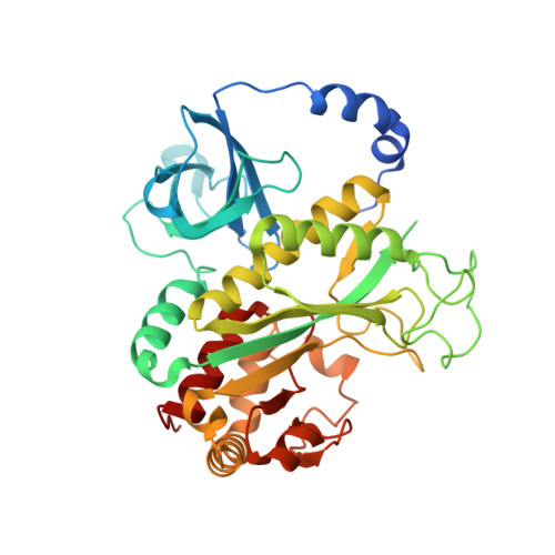
Crystal structures of mammalian glutamine synthetases illustrate substrate-induced conformational changes and provide opportunities for drug and herbicide design.

Krajewski, W.W.Collins, R.Holmberg-Schiavone, L.Jones, T.A.Karlberg, T.Mowbray, S.L.

(2008) J Mol Biol 375: 217-228

  • DOI: https://doi.org/10.1016/j.jmb.2007.10.029
  • Primary Citation of Related Structures:  
    2OJW, 2QC8, 2UU7

  • PubMed Abstract: 

    Glutamine synthetase (GS) catalyzes the ligation of glutamate and ammonia to form glutamine, with concomitant hydrolysis of ATP. In mammals, the activity eliminates cytotoxic ammonia, at the same time converting neurotoxic glutamate to harmless glutamine; there are a number of links between changes in GS activity and neurodegenerative disorders, such as Alzheimer's disease. In plants, because of its importance in the assimilation and re-assimilation of ammonia, the enzyme is a target of some herbicides. GS is also a central component of bacterial nitrogen metabolism and a potential drug target. Previous studies had investigated the structures of bacterial and plant GSs. In the present publication, we report the first structures of mammalian GSs. The apo form of the canine enzyme was solved by molecular replacement and refined at a resolution of 3 A. Two structures of human glutamine synthetase represent complexes with: a) phosphate, ADP, and manganese, and b) a phosphorylated form of the inhibitor methionine sulfoximine, ADP and manganese; these structures were refined to resolutions of 2.05 A and 2.6 A, respectively. Loop movements near the active site generate more closed forms of the eukaryotic enzymes when substrates are bound; the largest changes are associated with the binding of the nucleotide. Comparisons with earlier structures provide a basis for the design of drugs that are specifically directed at either human or bacterial enzymes. The site of binding the amino acid substrate is highly conserved in bacterial and eukaryotic GSs, whereas the nucleotide binding site varies to a much larger degree. Thus, the latter site offers the best target for specific drug design. Differences between mammalian and plant enzymes are much more subtle, suggesting that herbicides targeting GS must be designed with caution.


  • Organizational Affiliation

    Department of Cell and Molecular Biology, Biomedical Center, Uppsala University, Box 596, SE-751 24 Uppsala, Sweden.


Macromolecules
Find similar proteins by:  (by identity cutoff)  |  3D Structure
Entity ID: 1
MoleculeChains Sequence LengthOrganismDetailsImage
Glutamine synthetase
A, B, C, D, E
384Homo sapiensMutation(s): 0 
Gene Names: GLULGLNS
EC: 6.3.1.2
UniProt & NIH Common Fund Data Resources
Find proteins for P15104 (Homo sapiens)
Explore P15104 
Go to UniProtKB:  P15104
PHAROS:  P15104
GTEx:  ENSG00000135821 
Entity Groups  
Sequence Clusters30% Identity50% Identity70% Identity90% Identity95% Identity100% Identity
UniProt GroupP15104
Sequence Annotations
Expand
  • Reference Sequence
Small Molecules
Ligands 5 Unique
IDChains Name / Formula / InChI Key2D Diagram3D Interactions
ADP
Query on ADP

Download Ideal Coordinates CCD File 
CA [auth C],
LA [auth D],
M [auth A],
SA [auth E],
U [auth B]
ADENOSINE-5'-DIPHOSPHATE
C10 H15 N5 O10 P2
XTWYTFMLZFPYCI-KQYNXXCUSA-N
PO4
Query on PO4

Download Ideal Coordinates CCD File 
AA [auth C],
J [auth A],
JA [auth D],
RA [auth E],
S [auth B]
PHOSPHATE ION
O4 P
NBIIXXVUZAFLBC-UHFFFAOYSA-K
GOL
Query on GOL

Download Ideal Coordinates CCD File 
DA [auth C]
EA [auth C]
MA [auth D]
N [auth A]
TA [auth E]
DA [auth C],
EA [auth C],
MA [auth D],
N [auth A],
TA [auth E],
V [auth B]
GLYCEROL
C3 H8 O3
PEDCQBHIVMGVHV-UHFFFAOYSA-N
MN
Query on MN

Download Ideal Coordinates CCD File 
F [auth A]
FA [auth D]
G [auth A]
GA [auth D]
H [auth A]
F [auth A],
FA [auth D],
G [auth A],
GA [auth D],
H [auth A],
HA [auth D],
I [auth A],
IA [auth D],
NA [auth E],
O [auth B],
OA [auth E],
P [auth B],
PA [auth E],
Q [auth B],
QA [auth E],
R [auth B],
W [auth C],
X [auth C],
Y [auth C],
Z [auth C]
MANGANESE (II) ION
Mn
WAEMQWOKJMHJLA-UHFFFAOYSA-N
CL
Query on CL

Download Ideal Coordinates CCD File 
BA [auth C],
K [auth A],
KA [auth D],
L [auth A],
T [auth B]
CHLORIDE ION
Cl
VEXZGXHMUGYJMC-UHFFFAOYSA-M
Experimental Data & Validation

Experimental Data

  • Method: X-RAY DIFFRACTION
  • Resolution: 2.05 Å
  • R-Value Free: 0.212 
  • R-Value Work: 0.159 
  • R-Value Observed: 0.162 
  • Space Group: C 1 2 1
Unit Cell:
Length ( Å )Angle ( ˚ )
a = 177.6α = 90
b = 122.6β = 130.6
c = 126.6γ = 90
Software Package:
Software NamePurpose
REFMACrefinement
XDSdata reduction
XSCALEdata scaling
MOLREPphasing

Structure Validation

View Full Validation Report


Ligand Structure Quality Assessment 


Entry History 

Revision History  (Full details and data files)

  • Version 1.0: 2007-03-13
    Type: Initial release
  • Version 1.1: 2007-11-20
    Changes: Version format compliance
  • Version 1.2: 2011-07-13
    Changes: Advisory, Derived calculations, Version format compliance
  • Version 1.3: 2023-08-30
    Changes: Data collection, Database references, Derived calculations, Refinement description