2OHU

X-ray crystal structure of beta secretase complexed with compound 8b


Experimental Data Snapshot

  • Method: X-RAY DIFFRACTION
  • Resolution: 2.35 Å
  • R-Value Free: 0.248 
  • R-Value Work: 0.195 
  • R-Value Observed: 0.198 

wwPDB Validation   3D Report Full Report


Ligand Structure Quality Assessment 


This is version 1.5 of the entry. See complete history


Literature

Application of fragment screening by X-ray crystallography to the discovery of aminopyridines as inhibitors of beta-secretase.

Congreve, M.Aharony, D.Albert, J.Callaghan, O.Campbell, J.Carr, R.A.Chessari, G.Cowan, S.Edwards, P.D.Frederickson, M.McMenamin, R.Murray, C.W.Patel, S.Wallis, N.

(2007) J Med Chem 50: 1124-1132

  • DOI: https://doi.org/10.1021/jm061197u
  • Primary Citation of Related Structures:  
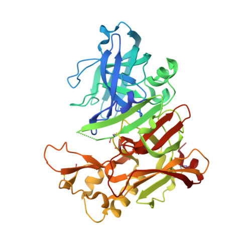
    2OHP, 2OHQ, 2OHR, 2OHS, 2OHT, 2OHU

  • PubMed Abstract: 

    Fragment-based lead discovery has been successfully applied to the aspartyl protease enzyme beta-secretase (BACE-1). Fragment hits that contained an aminopyridine motif binding to the two catalytic aspartic acid residues in the active site of the enzyme were the chemical starting points. Structure-based design approaches have led to identification of low micromolar lead compounds that retain these interactions and additionally occupy adjacent hydrophobic pockets of the active site. These leads form two subseries, for which compounds 4 (IC50 = 25 microM) and 6c (IC50 = 24 microM) are representative. In the latter series, further optimization has led to 8a (IC50 = 690 nM).


  • Organizational Affiliation

    Astex Therapeutics Ltd., 436 Cambridge Science Park, Milton Road, Cambridge CB4 0QA, United Kingdom. m.congreve@astex-therapeutics.com


Macromolecules
Find similar proteins by:  (by identity cutoff)  |  3D Structure
Entity ID: 1
MoleculeChains Sequence LengthOrganismDetailsImage
Beta-secretase 1402Homo sapiensMutation(s): 2 
Gene Names: BACE1BACE
EC: 3.4.23.46
UniProt & NIH Common Fund Data Resources
Find proteins for P56817 (Homo sapiens)
Explore P56817 
Go to UniProtKB:  P56817
PHAROS:  P56817
GTEx:  ENSG00000186318 
Entity Groups  
Sequence Clusters30% Identity50% Identity70% Identity90% Identity95% Identity100% Identity
UniProt GroupP56817
Sequence Annotations
Expand
  • Reference Sequence
Small Molecules
Binding Affinity Annotations 
IDSourceBinding Affinity
IP7 Binding MOAD:  2OHU IC50: 4200 (nM) from 1 assay(s)
PDBBind:  2OHU IC50: 4200 (nM) from 1 assay(s)
BindingDB:  2OHU IC50: 4200 (nM) from 1 assay(s)
Experimental Data & Validation

Experimental Data

  • Method: X-RAY DIFFRACTION
  • Resolution: 2.35 Å
  • R-Value Free: 0.248 
  • R-Value Work: 0.195 
  • R-Value Observed: 0.198 
  • Space Group: P 61 2 2
Unit Cell:
Length ( Å )Angle ( ˚ )
a = 102.162α = 90
b = 102.162β = 90
c = 168.385γ = 120
Software Package:
Software NamePurpose
REFMACrefinement
PDB_EXTRACTdata extraction
CrystalCleardata collection
MOSFLMdata reduction
CCP4data scaling
C-SEARCHphasing

Structure Validation

View Full Validation Report


Ligand Structure Quality Assessment 


Entry History 

Deposition Data

  • Released Date: 2007-03-13 
  • Deposition Author(s): Patel, S.

Revision History  (Full details and data files)

  • Version 1.0: 2007-03-13
    Type: Initial release
  • Version 1.1: 2008-05-01
    Changes: Version format compliance
  • Version 1.2: 2011-07-13
    Changes: Version format compliance
  • Version 1.3: 2017-10-18
    Changes: Advisory, Refinement description
  • Version 1.4: 2021-10-20
    Changes: Advisory, Database references, Derived calculations
  • Version 1.5: 2023-08-30
    Changes: Data collection, Refinement description