2OHO

Structural Basis for Glutamate Racemase Inhibitor


Experimental Data Snapshot

  • Method: X-RAY DIFFRACTION
  • Resolution: 2.25 Å
  • R-Value Free: 0.213 
  • R-Value Work: 0.207 
  • R-Value Observed: 0.207 

wwPDB Validation   3D Report Full Report


This is version 1.2 of the entry. See complete history


Literature

Structural basis for glutamate racemase inhibition

Kim, K.H.Bong, Y.J.Park, J.K.Shin, K.J.Hwang, K.Y.Kim, E.E.

(2007) J Mol Biol 372: 434-443

  • DOI: https://doi.org/10.1016/j.jmb.2007.05.003
  • Primary Citation of Related Structures:  
    2OHG, 2OHO, 2OHV

  • PubMed Abstract: 

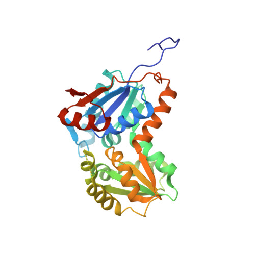
    D-Glutamic acid is a required biosynthetic building block for peptidoglycan, and the enzyme glutamate racemase (GluR) catalyzes the inter-conversion of D and L-glutamate enantiomers. Therefore, GluR is considered as an attractive target for the design of new antibacterial drugs. Here, we report the crystal structures of GluR from Streptococcus pyogenes in both inhibitor-free and inhibitor-bound forms. The inhibitor free GluR crystallized in two different forms, which diffracted to 2.25 A and 2.5 A resolution, while the inhibitor-bound crystal diffracted to 2.5 A resolution. GluR is composed of two domains of alpha/beta protein that are related by pseudo-2-fold symmetry and the active site is located at the domain interface. The inhibitor, gamma-2-naphthylmethyl-D-glutamate, which was reported earlier as a novel potent competitive inhibitor, makes several hydrogen bonds with protein atoms, and the naphthyl moiety is located in the hydrophobic pocket. The inhibitor binding induces a disorder in one of the loops near the active site. In both crystal forms, GluR exists as a dimer and the interactions seen at the dimer interface are almost identical. This agrees well with the results from gel filtration and dynamic light-scattering studies.


  • Organizational Affiliation

    Life Sciences Division, Korea Institute of Science and Technology, 39-1 Hawolkok-Dong, Sungbuk-Gu, Seoul, Korea.


Macromolecules
Find similar proteins by:  (by identity cutoff)  |  3D Structure
Entity ID: 1
MoleculeChains Sequence LengthOrganismDetailsImage
Glutamate Racemase
A, B
273Streptococcus pyogenes M1 GASMutation(s): 0 
Gene Names: murI
EC: 5.1.1.3
UniProt
Find proteins for Q9A1B7 (Streptococcus pyogenes serotype M1)
Explore Q9A1B7 
Go to UniProtKB:  Q9A1B7
Entity Groups  
Sequence Clusters30% Identity50% Identity70% Identity90% Identity95% Identity100% Identity
UniProt GroupQ9A1B7
Sequence Annotations
Expand
  • Reference Sequence
Experimental Data & Validation

Experimental Data

  • Method: X-RAY DIFFRACTION
  • Resolution: 2.25 Å
  • R-Value Free: 0.213 
  • R-Value Work: 0.207 
  • R-Value Observed: 0.207 
  • Space Group: P 21 21 21
Unit Cell:
Length ( Å )Angle ( ˚ )
a = 66.397α = 90
b = 88.951β = 90
c = 95.107γ = 90
Software Package:
Software NamePurpose
CNSrefinement
HKL-2000data collection
HKL-2000data reduction
SCALEPACKdata scaling
CNSphasing

Structure Validation

View Full Validation Report


Entry History 

Deposition Data

  • Released Date: 2007-09-25 
  • Deposition Author(s): Kim, E.E.

Revision History  (Full details and data files)

  • Version 1.0: 2007-09-25
    Type: Initial release
  • Version 1.1: 2011-07-13
    Changes: Version format compliance
  • Version 1.2: 2023-10-25
    Changes: Data collection, Database references, Refinement description