


Ligand Protection and Nitric Oxide Interactions with Heme in Nitrophorin 4: Kinetic and Structural Analyses of a Loop Mutant
Amoia, A.M., Montfort, W.R.To be published.
Experimental Data Snapshot
Entity ID: 1 | |||||
|---|---|---|---|---|---|
| Molecule | Chains | Sequence Length | Organism | Details | Image |
| Nitrophorin-4 | A [auth X] | 184 | Rhodnius prolixus | Mutation(s): 3 | 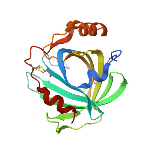 |
UniProt | |||||
Find proteins for Q94734 (Rhodnius prolixus) Explore Q94734 Go to UniProtKB: Q94734 | |||||
Entity Groups | |||||
| Sequence Clusters | 30% Identity50% Identity70% Identity90% Identity95% Identity100% Identity | ||||
| UniProt Group | Q94734 | ||||
Sequence AnnotationsExpand | |||||
| |||||
| Ligands 3 Unique | |||||
|---|---|---|---|---|---|
| ID | Chains | Name / Formula / InChI Key | 2D Diagram | 3D Interactions | |
| HEM Query on HEM | C [auth X] | PROTOPORPHYRIN IX CONTAINING FE C34 H32 Fe N4 O4 KABFMIBPWCXCRK-RGGAHWMASA-L |  | ||
| PO4 Query on PO4 | B [auth X] | PHOSPHATE ION O4 P NBIIXXVUZAFLBC-UHFFFAOYSA-K |  | ||
| NO Query on NO | D [auth X] | NITRIC OXIDE N O ODUCDPQEXGNKDN-UHFFFAOYSA-N |  | ||
| Length ( Å ) | Angle ( ˚ ) |
|---|---|
| a = 70.191 | α = 90 |
| b = 42.637 | β = 94.12 |
| c = 52.975 | γ = 90 |
| Software Name | Purpose |
|---|---|
| REFMAC | refinement |
| CrystalClear | data scaling |
RCSB PDB (citation) is hosted by
RCSB PDB is a member of the

