2OF3

TOG domain structure from C.elegans Zyg9


Experimental Data Snapshot

  • Method: X-RAY DIFFRACTION
  • Resolution: 1.90 Å
  • R-Value Free: 0.224 
  • R-Value Work: 0.173 
  • R-Value Observed: 0.175 

wwPDB Validation   3D Report Full Report


This is version 1.3 of the entry. See complete history


Literature

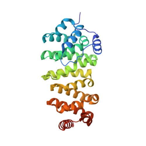
Crystal structure of a TOG domain: conserved features of XMAP215/Dis1-family TOG domains and implications for tubulin binding.

Al-Bassam, J.Larsen, N.A.Hyman, A.A.Harrison, S.C.

(2007) Structure 15: 355-362

  • DOI: https://doi.org/10.1016/j.str.2007.01.012
  • Primary Citation of Related Structures:  
    2OF3

  • PubMed Abstract: 

    Members of the XMAP215/Dis1 family of microtubule-associated proteins (MAPs) are essential for microtubule growth. MAPs in this family contain several 250 residue repeats, called TOG domains, which are thought to bind tubulin dimers and promote microtubule polymerization. We have determined the crystal structure of a single TOG domain from the Caenorhabditis elegans homolog, Zyg9, to 1.9 A resolution, and from it we describe a structural blueprint for TOG domains. These domains are flat, paddle-like structures, composed of six HEAT-repeat elements stacked side by side. The two wide faces of the paddle contain the HEAT-repeat helices, and the two narrow faces, the intra- and inter-HEAT repeat turns. Solvent-exposed residues in the intrarepeat turns are conserved, both within a particular protein and across the XMAP215/Dis1 family. Mutation of some of these residues in the TOG1 domain from the budding yeast homolog, Stu2p, shows that this face indeed participates in the tubulin contact.


  • Organizational Affiliation

    Jack and Eileen Connors Laboratory of Structural Biology , Harvard Medical School, Boston, MA 02115, USA.


Macromolecules
Find similar proteins by:  (by identity cutoff)  |  3D Structure
Entity ID: 1
MoleculeChains Sequence LengthOrganismDetailsImage
ZYG-9266Caenorhabditis elegansMutation(s): 0 
Gene Names: zyg-9
UniProt
Find proteins for G5EEM5 (Caenorhabditis elegans)
Explore G5EEM5 
Go to UniProtKB:  G5EEM5
Entity Groups  
Sequence Clusters30% Identity50% Identity70% Identity90% Identity95% Identity100% Identity
UniProt GroupG5EEM5
Sequence Annotations
Expand
  • Reference Sequence
Experimental Data & Validation

Experimental Data

  • Method: X-RAY DIFFRACTION
  • Resolution: 1.90 Å
  • R-Value Free: 0.224 
  • R-Value Work: 0.173 
  • R-Value Observed: 0.175 
  • Space Group: P 41
Unit Cell:
Length ( Å )Angle ( ˚ )
a = 54.13α = 90
b = 54.13β = 90
c = 116.53γ = 90
Software Package:
Software NamePurpose
REFMACrefinement
ADSCdata collection
HKL-2000data reduction
HKL-2000data scaling
SOLVEphasing

Structure Validation

View Full Validation Report


Entry History 

Deposition Data

Revision History  (Full details and data files)

  • Version 1.0: 2007-03-13
    Type: Initial release
  • Version 1.1: 2008-05-01
    Changes: Version format compliance
  • Version 1.2: 2011-07-13
    Changes: Version format compliance
  • Version 1.3: 2023-12-27
    Changes: Data collection, Database references