2ODR

Methanococcus Maripaludis Phosphoseryl-tRNA synthetase


Experimental Data Snapshot

  • Method: X-RAY DIFFRACTION
  • Resolution: 3.23 Å
  • R-Value Free: 0.306 
  • R-Value Work: 0.292 
  • R-Value Observed: 0.292 

wwPDB Validation   3D Report Full Report


This is version 1.4 of the entry. See complete history


Literature

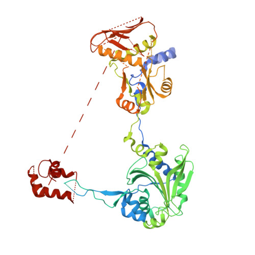
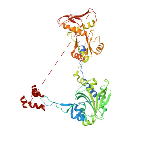
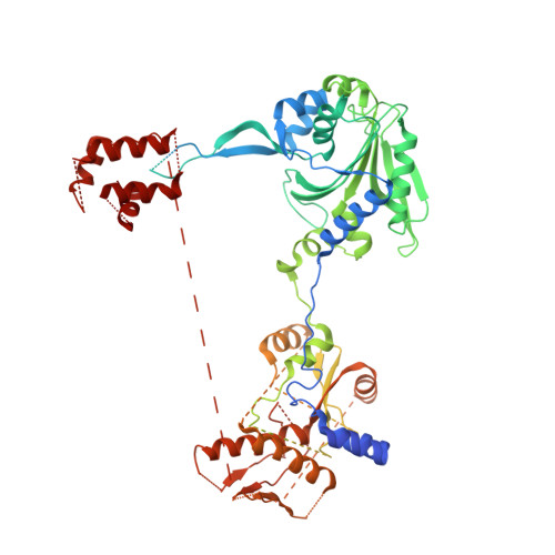
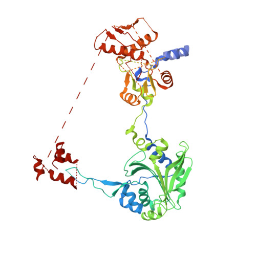
Toward understanding phosphoseryl-tRNACys formation: the crystal structure of Methanococcus maripaludis phosphoseryl-tRNA synthetase.

Kamtekar, S.Hohn, M.J.Park, H.S.Schnitzbauer, M.Sauerwald, A.Soll, D.Steitz, T.A.

(2007) Proc Natl Acad Sci U S A 104: 2620-2625

  • DOI: https://doi.org/10.1073/pnas.0611504104
  • Primary Citation of Related Structures:  
    2ODR

  • PubMed Abstract: 

    A number of archaeal organisms generate Cys-tRNA(Cys) in a two-step pathway, first charging phosphoserine (Sep) onto tRNA(Cys) and subsequently converting it to Cys-tRNA(Cys). We have determined, at 3.2-A resolution, the structure of the Methanococcus maripaludis phosphoseryl-tRNA synthetase (SepRS), which catalyzes the first step of this pathway. The structure shows that SepRS is a class II, alpha(4) synthetase whose quaternary structure arrangement of subunits closely resembles that of the heterotetrameric (alphabeta)(2) phenylalanyl-tRNA synthetase (PheRS). Homology modeling of a tRNA complex indicates that, in contrast to PheRS, a single monomer in the SepRS tetramer may recognize both the acceptor terminus and anticodon of a tRNA substrate. Using a complex with tungstate as a marker for the position of the phosphate moiety of Sep, we suggest that SepRS and PheRS bind their respective amino acid substrates in dissimilar orientations by using different residues.


  • Organizational Affiliation

    Department of Molecular Biophysics and Biochemistry and Chemistry and Howard Hughes Medical Institute, Yale University, New Haven, CT 06520-8114, USA.


Macromolecules
Find similar proteins by:  (by identity cutoff)  |  3D Structure
Entity ID: 1
MoleculeChains Sequence LengthOrganismDetailsImage
phosphoseryl-tRNA synthetase665Methanococcus maripaludis S2Mutation(s): 0 
Gene Names: MMP0688
EC: 6.1.1
UniProt
Find proteins for Q6LZE1 (Methanococcus maripaludis (strain S2 / LL))
Explore Q6LZE1 
Go to UniProtKB:  Q6LZE1
Entity Groups  
Sequence Clusters30% Identity50% Identity70% Identity90% Identity95% Identity100% Identity
UniProt GroupQ6LZE1
Sequence Annotations
Expand
  • Reference Sequence
Find similar proteins by:  (by identity cutoff)  |  3D Structure
Entity ID: 2
MoleculeChains Sequence LengthOrganismDetailsImage
phosphoseryl-tRNA synthetase648Methanococcus maripaludis S2Mutation(s): 0 
Gene Names: MMP0688
EC: 6.1.1
UniProt
Find proteins for Q6LZE1 (Methanococcus maripaludis (strain S2 / LL))
Explore Q6LZE1 
Go to UniProtKB:  Q6LZE1
Entity Groups  
Sequence Clusters30% Identity50% Identity70% Identity90% Identity95% Identity100% Identity
UniProt GroupQ6LZE1
Sequence Annotations
Expand
  • Reference Sequence
Find similar proteins by:  (by identity cutoff)  |  3D Structure
Entity ID: 3
MoleculeChains Sequence LengthOrganismDetailsImage
phosphoseryl-tRNA synthetase701Methanococcus maripaludis S2Mutation(s): 0 
Gene Names: MMP0688
EC: 6.1.1
UniProt
Find proteins for Q6LZE1 (Methanococcus maripaludis (strain S2 / LL))
Explore Q6LZE1 
Go to UniProtKB:  Q6LZE1
Entity Groups  
Sequence Clusters30% Identity50% Identity70% Identity90% Identity95% Identity100% Identity
UniProt GroupQ6LZE1
Sequence Annotations
Expand
  • Reference Sequence
Find similar proteins by:  (by identity cutoff)  |  3D Structure
Entity ID: 4
MoleculeChains Sequence LengthOrganismDetailsImage
phosphoseryl-tRNA synthetase685Methanococcus maripaludis S2Mutation(s): 0 
Gene Names: MMP0688
EC: 6.1.1
UniProt
Find proteins for Q6LZE1 (Methanococcus maripaludis (strain S2 / LL))
Explore Q6LZE1 
Go to UniProtKB:  Q6LZE1
Entity Groups  
Sequence Clusters30% Identity50% Identity70% Identity90% Identity95% Identity100% Identity
UniProt GroupQ6LZE1
Sequence Annotations
Expand
  • Reference Sequence
Experimental Data & Validation

Experimental Data

  • Method: X-RAY DIFFRACTION
  • Resolution: 3.23 Å
  • R-Value Free: 0.306 
  • R-Value Work: 0.292 
  • R-Value Observed: 0.292 
  • Space Group: P 21 21 21
Unit Cell:
Length ( Å )Angle ( ˚ )
a = 119.172α = 90
b = 133.943β = 90
c = 208.382γ = 90
Software Package:
Software NamePurpose
DENZOdata reduction
SCALEPACKdata scaling
SOLVEphasing
REFMACrefinement
PDB_EXTRACTdata extraction
ADSCdata collection
HKL-2000data reduction

Structure Validation

View Full Validation Report


Entry History 

Deposition Data

Revision History  (Full details and data files)

  • Version 1.0: 2007-02-13
    Type: Initial release
  • Version 1.1: 2008-05-01
    Changes: Version format compliance
  • Version 1.2: 2011-07-13
    Changes: Advisory, Source and taxonomy, Version format compliance
  • Version 1.3: 2017-10-18
    Changes: Refinement description
  • Version 1.4: 2023-12-27
    Changes: Data collection, Database references, Refinement description