2ODP

Complement component C2a, the catalytic fragment of C3- and C5-convertase of human complement


Experimental Data Snapshot

  • Method: X-RAY DIFFRACTION
  • Resolution: 1.90 Å
  • R-Value Free: 0.250 
  • R-Value Work: 0.207 
  • R-Value Observed: 0.209 

wwPDB Validation   3D Report Full Report


Ligand Structure Quality Assessment 


This is version 2.2 of the entry. See complete history


Literature

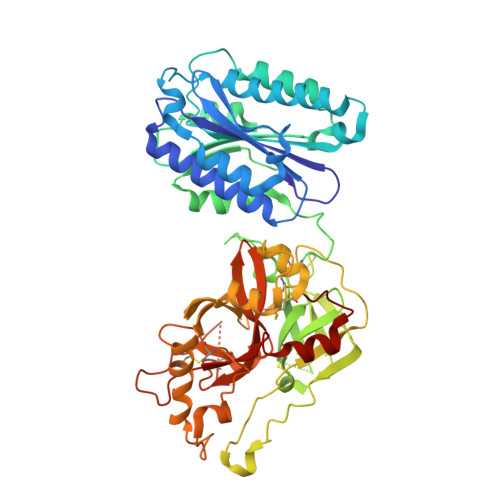
The crystal structure of c2a, the catalytic fragment of classical pathway c3 and c5 convertase of human complement.

Krishnan, V.Xu, Y.Macon, K.Volanakis, J.E.Narayana, S.V.

(2007) J Mol Biol 367: 224-233

  • DOI: https://doi.org/10.1016/j.jmb.2006.12.039
  • Primary Citation of Related Structures:  
    2ODP, 2ODQ

  • PubMed Abstract: 

    The multi-domain serine protease C2 provides the catalytic activity for the C3 and C5- convertases of the classical and lectin pathways of complement activation. Formation of these convertases requires the Mg(2+)-dependent binding of C2 to C4b, and the subsequent cleavage of C2 by C1s or MASP2, respectively. The C-terminal fragment C2a consisting of a serine protease (SP) and a von Willebrand factor type A (vWFA) domain, remains attached to C4b, forming the C3 convertase, C4b2a. Here, we present the crystal structure of Mg(2+)-bound C2a to 1.9 A resolution in comparison to its homolog Bb, the catalytic subunit of the alternative pathway C3 convertase, C3bBb. Although the overall domain arrangement of C2a is similar to Bb, there are certain structural differences. Unexpectedly, the conformation of the metal ion-dependent adhesion site and the position of the alpha7 helix of the vWFA domain indicate a co-factor-bound or open conformation. The active site of the SP domain is in a zymogen-like inactive conformation. On the basis of these structural features, we suggest a model for the initial steps of C3 convertase assembly.


  • Organizational Affiliation

    Center for Biophysical Sciences and Engineering, School of Optometry, University of Alabama at Birmingham, Birmingham, AL 35294, USA.


Macromolecules
Find similar proteins by:  (by identity cutoff)  |  3D Structure
Entity ID: 1
MoleculeChains Sequence LengthOrganismDetailsImage
Complement C2509Homo sapiensMutation(s): 1 
EC: 3.4.21.43
UniProt
Find proteins for Q5JP69 (Homo sapiens)
Explore Q5JP69 
Go to UniProtKB:  Q5JP69
Entity Groups  
Sequence Clusters30% Identity50% Identity70% Identity90% Identity95% Identity100% Identity
UniProt GroupQ5JP69
Sequence Annotations
Expand
  • Reference Sequence
Oligosaccharides

Help

Entity ID: 2
MoleculeChains Length2D Diagram Glycosylation3D Interactions
2-acetamido-2-deoxy-beta-D-glucopyranose-(1-3)-[2-acetamido-2-deoxy-beta-D-glucopyranose-(1-4)]2-acetamido-2-deoxy-beta-D-glucopyranose
B
3N-Glycosylation
Glycosylation Resources
GlyTouCan:  G26744ZJ
GlyCosmos:  G26744ZJ
GlyGen:  G26744ZJ
Entity ID: 3
MoleculeChains Length2D Diagram Glycosylation3D Interactions
2-acetamido-2-deoxy-beta-D-glucopyranose-(1-4)-2-acetamido-2-deoxy-beta-D-glucopyranose-(1-4)-2-acetamido-2-deoxy-beta-D-glucopyranose
C
3N-Glycosylation
Glycosylation Resources
GlyTouCan:  G47362BJ
GlyCosmos:  G47362BJ
GlyGen:  G47362BJ
Biologically Interesting Molecules (External Reference) 1 Unique
Experimental Data & Validation

Experimental Data

  • Method: X-RAY DIFFRACTION
  • Resolution: 1.90 Å
  • R-Value Free: 0.250 
  • R-Value Work: 0.207 
  • R-Value Observed: 0.209 
  • Space Group: P 1 21 1
Unit Cell:
Length ( Å )Angle ( ˚ )
a = 51.398α = 90
b = 83.819β = 92.1
c = 75.243γ = 90
Software Package:
Software NamePurpose
REFMACrefinement
PDB_EXTRACTdata extraction
HKL-2000data reduction
HKL-2000data scaling
CNSphasing

Structure Validation

View Full Validation Report


Ligand Structure Quality Assessment 


Entry History 

Deposition Data

Revision History  (Full details and data files)

  • Version 1.0: 2007-02-06
    Type: Initial release
  • Version 1.1: 2008-05-01
    Changes: Version format compliance
  • Version 1.2: 2011-07-13
    Changes: Non-polymer description, Version format compliance
  • Version 2.0: 2020-07-29
    Type: Remediation
    Reason: Carbohydrate remediation
    Changes: Atomic model, Data collection, Derived calculations, Structure summary
  • Version 2.1: 2021-11-10
    Changes: Database references, Structure summary
  • Version 2.2: 2023-10-25
    Changes: Data collection, Refinement description