2OD3

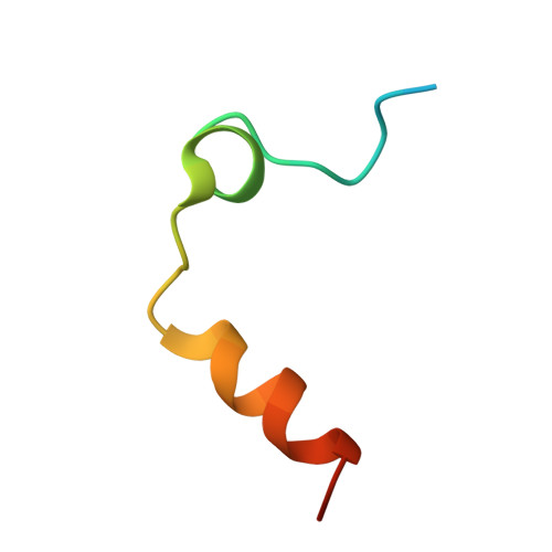
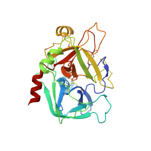
Human thrombin chimera with human residues 184a, 186, 186a, 186b, 186c and 222 replaced by murine thrombin equivalents.


Experimental Data Snapshot

  • Method: X-RAY DIFFRACTION
  • Resolution: 1.75 Å
  • R-Value Free: 0.230 
  • R-Value Work: 0.199 
  • R-Value Observed: 0.199 

wwPDB Validation   3D Report Full Report


Ligand Structure Quality Assessment 


This is version 1.7 of the entry. See complete history


Literature

Structural basis of na+ activation mimicry in murine thrombin.

Marino, F.Chen, Z.W.Ergenekan, C.E.Bush-Pelc, L.A.Mathews, F.S.Di Cera, E.

(2007) J Biol Chem 282: 16355-16361

  • DOI: https://doi.org/10.1074/jbc.M701323200
  • Primary Citation of Related Structures:  
    2OCV, 2OD3

  • PubMed Abstract: 

    Unlike human thrombin, murine thrombin lacks Na+ activation due to the charge reversal substitution D222K in the Na+ binding loop. However, the enzyme is functionally stabilized in a Na+-bound form and is highly active toward physiologic substrates. The structural basis of this peculiar property is unknown. Here, we present the 2.2 A resolution x-ray crystal structure of murine thrombin in the absence of inhibitors and salts. The enzyme assumes an active conformation, with Ser-195, Glu-192, and Asp-189 oriented as in the Na+-bound fast form of human thrombin. Lys-222 completely occludes the pore of entry to the Na+ binding site and positions its side chain inside the pore, with the Nzeta atom H-bonded to the backbone oxygen atoms of Lys-185, Asp-186b, and Lys-186d. The same architecture is observed in the 1.75 A resolution structure of a thrombin chimera in which the human enzyme carries all residues defining the Na+ pore in the murine enzyme. These findings demonstrate that Na+ activation in thrombin is linked to the architecture of the Na+ pore. The molecular strategy of Na+ activation mimicry unraveled for murine thrombin is relevant to serine proteases and enzymes activated by monovalent cations in general.


  • Organizational Affiliation

    Department of Biochemistry and Molecular Biophysics, Washington University Medical School, St. Louis, Missouri 63110, USA.


Macromolecules
Find similar proteins by:  (by identity cutoff)  |  3D Structure
Entity ID: 1
MoleculeChains Sequence LengthOrganismDetailsImage
Thrombin light chain36Homo sapiensMutation(s): 0 
Gene Names: F2
UniProt & NIH Common Fund Data Resources
Find proteins for P00734 (Homo sapiens)
Explore P00734 
Go to UniProtKB:  P00734
PHAROS:  P00734
GTEx:  ENSG00000180210 
Entity Groups  
Sequence Clusters30% Identity50% Identity70% Identity90% Identity95% Identity100% Identity
UniProt GroupP00734
Sequence Annotations
Expand
  • Reference Sequence
Find similar proteins by:  (by identity cutoff)  |  3D Structure
Entity ID: 2
MoleculeChains Sequence LengthOrganismDetailsImage
Thrombin heavy chain259Homo sapiensMutation(s): 6 
Gene Names: F2
UniProt & NIH Common Fund Data Resources
Find proteins for P00734 (Homo sapiens)
Explore P00734 
Go to UniProtKB:  P00734
PHAROS:  P00734
GTEx:  ENSG00000180210 
Entity Groups  
Sequence Clusters30% Identity50% Identity70% Identity90% Identity95% Identity100% Identity
UniProt GroupP00734
Sequence Annotations
Expand
  • Reference Sequence
Small Molecules
Ligands 2 Unique
IDChains Name / Formula / InChI Key2D Diagram3D Interactions
0G6
Query on 0G6

Download Ideal Coordinates CCD File 
C [auth B]D-phenylalanyl-N-[(2S,3S)-6-{[amino(iminio)methyl]amino}-1-chloro-2-hydroxyhexan-3-yl]-L-prolinamide
C21 H34 Cl N6 O3
DVFLYEYCMMLBTQ-VSZNYVQBSA-O
NAG
Query on NAG

Download Ideal Coordinates CCD File 
D [auth B]2-acetamido-2-deoxy-beta-D-glucopyranose
C8 H15 N O6
OVRNDRQMDRJTHS-FMDGEEDCSA-N
Biologically Interesting Molecules (External Reference) 1 Unique
Experimental Data & Validation

Experimental Data

  • Method: X-RAY DIFFRACTION
  • Resolution: 1.75 Å
  • R-Value Free: 0.230 
  • R-Value Work: 0.199 
  • R-Value Observed: 0.199 
  • Space Group: P 21 21 21
Unit Cell:
Length ( Å )Angle ( ˚ )
a = 44.952α = 90
b = 74.078β = 90
c = 103.565γ = 90
Software Package:
Software NamePurpose
CNSrefinement
ADSCdata collection
HKL-2000data reduction
SCALEPACKdata scaling
CCP4phasing

Structure Validation

View Full Validation Report


Ligand Structure Quality Assessment 


Entry History 

Deposition Data

Revision History  (Full details and data files)

  • Version 1.0: 2007-04-10
    Type: Initial release
  • Version 1.1: 2008-05-01
    Changes: Version format compliance
  • Version 1.2: 2011-07-13
    Changes: Atomic model, Database references, Derived calculations, Non-polymer description, Structure summary, Version format compliance
  • Version 1.3: 2013-02-27
    Changes: Other
  • Version 1.4: 2020-07-29
    Type: Remediation
    Reason: Carbohydrate remediation
    Changes: Data collection, Derived calculations, Structure summary
  • Version 1.5: 2021-10-20
    Changes: Database references, Structure summary
  • Version 1.6: 2023-08-30
    Changes: Data collection, Refinement description
  • Version 1.7: 2024-03-13
    Changes: Source and taxonomy, Structure summary