2OC2

Structure of testis ACE with RXPA380


Experimental Data Snapshot

  • Method: X-RAY DIFFRACTION
  • Resolution: 2.25 Å
  • R-Value Free: 0.262 
  • R-Value Work: 0.215 
  • R-Value Observed: 0.217 

wwPDB Validation   3D Report Full Report


Ligand Structure Quality Assessment 


This is version 1.6 of the entry. See complete history


Literature

The structure of testis angiotensin-converting enzyme in complex with the C domain-specific inhibitor RXPA380.

Corradi, H.R.Chitapi, I.Sewell, B.T.Georgiadis, D.Dive, V.Sturrock, E.D.Acharya, K.R.

(2007) Biochemistry 46: 5473-5478

  • DOI: https://doi.org/10.1021/bi700275e
  • Primary Citation of Related Structures:  
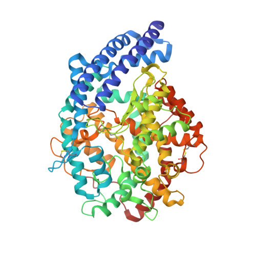
    2OC2

  • PubMed Abstract: 

    Angiotensin I-converting enzyme (ACE) is central to the regulation of the renin-angiotensin system and is a key therapeutic target for combating hypertension and related cardiovascular diseases. Currently available drugs bind both active sites of its two homologous domains, although it is now understood that these domains function differently in vivo. The recently solved crystal structures of both domains (N and C) open the door to new domain-specific inhibitor design, taking advantage of the differences between these two large active sites. Here we present the first crystal structure at a resolution of 2.25 A of testis ACE (identical to the C domain of somatic ACE) with the highly C-domain-specific phosphinic inhibitor, RXPA380. Testis ACE retains the same conformation as seen in previously determined inhibitor complexes, but the RXPA380 central backbone conformation is more similar to that seen for the inhibitor captopril than enalaprilat. The RXPA380 molecule occupies more subsites of the testis ACE active site than the previously determined inhibitors and possesses bulky moieties that extend into the S2' and S2 subsites. Thus the high affinity of RXPA380 for the testis ACE/somatic ACE C domain is explained by the interaction of these bulky moieties with residues unique to these domains, specifically Phe 391, Val 379, and Val 380, that are not found in the N domain. The characterization of the extended active site and the binding of a potent C-domain-selective inhibitor provide the first structural data for the design of truly domain-specific pharmacophores.


  • Organizational Affiliation

    Department of Biology and Biochemistry, University of Bath, Claverton Down, Bath BA2 7AY, United Kingdom.


Macromolecules
Find similar proteins by:  (by identity cutoff)  |  3D Structure
Entity ID: 1
MoleculeChains Sequence LengthOrganismDetailsImage
Angiotensin-converting enzyme, somatic isoform591Homo sapiensMutation(s): 0 
Gene Names: ACEDCPDCP1
EC: 3.4.15.1
UniProt & NIH Common Fund Data Resources
Find proteins for P12821 (Homo sapiens)
Explore P12821 
Go to UniProtKB:  P12821
PHAROS:  P12821
GTEx:  ENSG00000159640 
Entity Groups  
Sequence Clusters30% Identity50% Identity70% Identity90% Identity95% Identity100% Identity
UniProt GroupP12821
Sequence Annotations
Expand
  • Reference Sequence
Small Molecules
Binding Affinity Annotations 
IDSourceBinding Affinity
RX3 BindingDB:  2OC2 Ki: min: 3, max: 1.00e+4 (nM) from 3 assay(s)
PDBBind:  2OC2 Ki: 3 (nM) from 1 assay(s)
Binding MOAD:  2OC2 Ki: 3 (nM) from 1 assay(s)
Experimental Data & Validation

Experimental Data

  • Method: X-RAY DIFFRACTION
  • Resolution: 2.25 Å
  • R-Value Free: 0.262 
  • R-Value Work: 0.215 
  • R-Value Observed: 0.217 
  • Space Group: P 21 21 21
Unit Cell:
Length ( Å )Angle ( ˚ )
a = 56.514α = 90
b = 84.763β = 90
c = 133.483γ = 90
Software Package:
Software NamePurpose
REFMACrefinement
PDB_EXTRACTdata extraction
HKL-2000data collection
HKL-2000data reduction
SCALEPACKdata scaling

Structure Validation

View Full Validation Report


Ligand Structure Quality Assessment 


Entry History 

Deposition Data

Revision History  (Full details and data files)

  • Version 1.0: 2007-05-22
    Type: Initial release
  • Version 1.1: 2008-05-01
    Changes: Version format compliance
  • Version 1.2: 2011-07-13
    Changes: Version format compliance
  • Version 1.3: 2012-04-11
    Changes: Non-polymer description
  • Version 1.4: 2023-12-27
    Changes: Data collection, Database references, Derived calculations, Structure summary
  • Version 1.5: 2024-03-20
    Changes: Database references
  • Version 1.6: 2024-04-03
    Changes: Refinement description