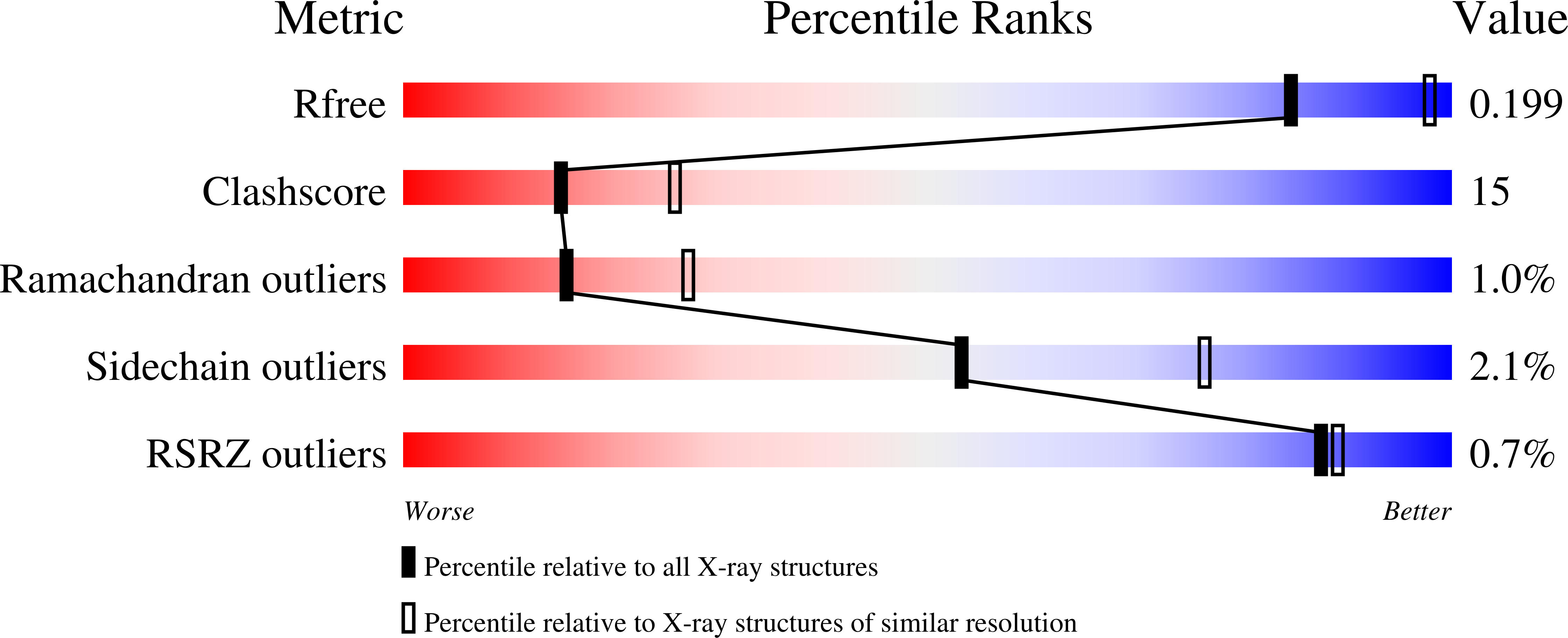
2OBX

Lumazine synthase RibH2 from Mesorhizobium loti (Gene mll7281, Swiss-Prot entry Q986N2) complexed with inhibitor 5-Nitro-6-(D-Ribitylamino)-2,4(1H,3H) Pyrimidinedione


Experimental Data Snapshot

  • Method: X-RAY DIFFRACTION
  • Resolution: 2.53 Å
  • R-Value Free: 0.281 
  • R-Value Work: 0.213 
  • R-Value Observed: 0.216 

wwPDB Validation   3D Report Full Report


Ligand Structure Quality Assessment 


This is version 1.4 of the entry. See complete history


Literature

Structural and kinetic properties of lumazine synthase isoenzymes in the order rhizobiales

Klinke, S.Zylberman, V.Bonomi, H.R.Haase, I.Guimaraes, B.G.Braden, B.C.Bacher, A.Fischer, M.Goldbaum, F.A.

(2007) J Mol Biol 373: 664-680

  • DOI: https://doi.org/10.1016/j.jmb.2007.08.021
  • Primary Citation of Related Structures:  
    2F59, 2I0F, 2O6H, 2OBX

  • PubMed Abstract: 

    6,7-Dimethyl-8-ribityllumazine synthase (lumazine synthase; LS) catalyzes the penultimate step in the biosynthesis of riboflavin in plants and microorganisms. This protein is known to exhibit different quaternary assemblies between species, existing as free pentamers, decamers (dimers of pentamers) and icosahedrally arranged dodecamers of pentamers. A phylogenetic analysis on eubacterial, fungal and plant LSs allowed us to classify them into two categories: Type I LSs (pentameric or icosahedral) and Type II LSs (decameric). The Rhizobiales represent an order of alpha-proteobacteria that includes, among others, the genera Mesorhizobium, Agrobacterium and Brucella. Here, we present structural and kinetic studies on several LSs from Rhizobiales. Interestingly, Mesorhizobium and Brucella encode both a Type-I LS and a Type-II LS called RibH1 and RibH2, respectively. We show that Type II LSs appear to be almost inactive, whereas Type I LSs present a highly variable catalytic activity according to the genus. Additionally, we have solved four RibH1/RibH2 crystallographic structures from the genera Mesorhizobium and Brucella. The relationship between the active-site architecture and catalytic properties in these isoenzymes is discussed, and a model that describes the enzymatic behavior is proposed. Furthermore, sequence alignment studies allowed us to extend our results to the genus Agrobacterium. Our results suggest that the selective pressure controlling the riboflavin pathway favored the evolution of catalysts with low reaction rates, since the excess of flavins in the intracellular pool in Rhizobiales could act as a negative factor when these bacteria are exposed to oxidative or nitrosative stress.


  • Organizational Affiliation

    Fundación Instituto Leloir, IIBBA-CONICET, C1405BWE, Buenos Aires, Argentina. sklinke@leloir.org.ar


Macromolecules
Find similar proteins by:  (by identity cutoff)  |  3D Structure
Entity ID: 1
MoleculeChains Sequence LengthOrganismDetailsImage
6,7-dimethyl-8-ribityllumazine synthase 1
A, B, C, D, E
A, B, C, D, E, F, G, H, I, J
157Mesorhizobium lotiMutation(s): 0 
Gene Names: ribH1ribH
EC: 2.5.1.78
UniProt
Find proteins for Q986N2 (Mesorhizobium japonicum (strain LMG 29417 / CECT 9101 / MAFF 303099))
Explore Q986N2 
Go to UniProtKB:  Q986N2
Entity Groups  
Sequence Clusters30% Identity50% Identity70% Identity90% Identity95% Identity100% Identity
UniProt GroupQ986N2
Sequence Annotations
Expand
  • Reference Sequence
Small Molecules
Ligands 2 Unique
IDChains Name / Formula / InChI Key2D Diagram3D Interactions
INI
Query on INI

Download Ideal Coordinates CCD File 
CA [auth J]
L [auth A]
N [auth B]
P [auth C]
Q [auth D]
CA [auth J],
L [auth A],
N [auth B],
P [auth C],
Q [auth D],
S [auth E],
U [auth F],
W [auth G],
X [auth H],
Z [auth I]
5-NITRO-6-RIBITYL-AMINO-2,4(1H,3H)-PYRIMIDINEDIONE
C9 H14 N4 O8
KSKGHNZSCSCHEQ-RPDRRWSUSA-N
PO4
Query on PO4

Download Ideal Coordinates CCD File 
AA [auth J]
BA [auth J]
K [auth A]
M [auth B]
O [auth C]
AA [auth J],
BA [auth J],
K [auth A],
M [auth B],
O [auth C],
R [auth E],
T [auth F],
V [auth G],
Y [auth I]
PHOSPHATE ION
O4 P
NBIIXXVUZAFLBC-UHFFFAOYSA-K
Experimental Data & Validation

Experimental Data

  • Method: X-RAY DIFFRACTION
  • Resolution: 2.53 Å
  • R-Value Free: 0.281 
  • R-Value Work: 0.213 
  • R-Value Observed: 0.216 
  • Space Group: C 1 2 1
Unit Cell:
Length ( Å )Angle ( ˚ )
a = 154.385α = 90
b = 122.218β = 125.43
c = 94.929γ = 90
Software Package:
Software NamePurpose
MAR345data collection
AMoREphasing
CNSrefinement
MOSFLMdata reduction
SCALAdata scaling

Structure Validation

View Full Validation Report


Ligand Structure Quality Assessment 


Entry History 

Deposition Data

Revision History  (Full details and data files)

  • Version 1.0: 2007-08-14
    Type: Initial release
  • Version 1.1: 2011-07-13
    Changes: Derived calculations, Version format compliance
  • Version 1.2: 2013-01-16
    Changes: Structure summary
  • Version 1.3: 2017-10-18
    Changes: Refinement description
  • Version 1.4: 2023-08-30
    Changes: Data collection, Database references, Derived calculations, Refinement description