2OAN

Structure of oxidized beta-actin


Experimental Data Snapshot

  • Method: X-RAY DIFFRACTION
  • Resolution: 2.61 Å
  • R-Value Free: 0.288 
  • R-Value Work: 0.210 
  • R-Value Observed: 0.214 

wwPDB Validation   3D Report Full Report


Ligand Structure Quality Assessment 


This is version 1.5 of the entry. See complete history


Literature

Molecular and Structural Basis for Redox Regulation of beta-Actin.

Lassing, I.Schmitzberger, F.Bjornstedt, M.Holmgren, A.Nordlund, P.Schutt, C.E.Lindberg, U.

(2007) J Mol Biol 370: 331-348

  • DOI: https://doi.org/10.1016/j.jmb.2007.04.056
  • Primary Citation of Related Structures:  
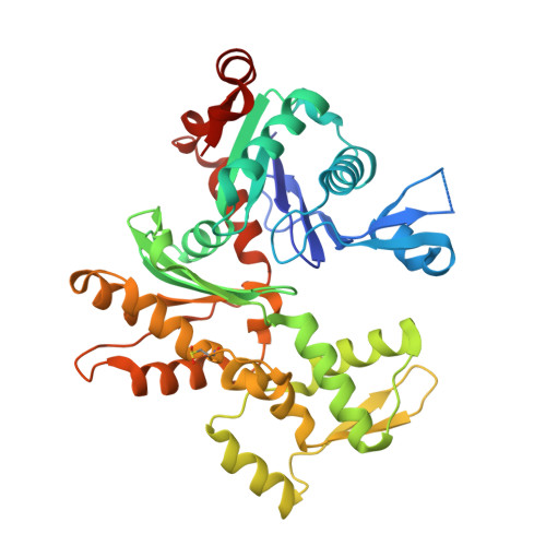
    2OAN

  • PubMed Abstract: 

    An essential consequence of growth factor-mediated signal transduction is the generation of intracellular H(2)O(2). It operates as a second messenger in the control of actin microfilament dynamics, causing rapid and dramatic changes in the morphology and motile activity of stimulated cells. Little is understood about the molecular mechanisms causing these changes in the actin system. Here, it is shown that H(2)O(2) acts directly upon several levels of this system, and some of the mechanistic effects are detailed. We describe the impact of oxidation on the polymerizability of non-muscle beta/gamma-actin and compare with that of muscle alpha-actin. Oxidation of beta/gamma-actin can cause a complete loss of polymerizability, crucially, reversible by the thioredoxin system. Further, oxidation of the actin impedes its interaction with profilin and causes depolymerization of filamentous actin. The effects of oxidation are critically dependent on the nucleotide state and the concentration of Ca(2+). We have determined the crystal structure of oxidized beta-actin to a resolution of 2.6 A. The arrangement in the crystal implies an antiparallel homodimer connected by an intermolecular disulfide bond involving cysteine 374. Our data indicate that this dimer forms under non-polymerizing and oxidizing conditions. We identify oxidation of cysteine 272 in the crystallized actin dimer, likely to a cysteine sulfinic acid. In beta/gamma-actin, this is the cysteine residue most reactive towards H(2)O(2) in solution, and we suggest plausible structural determinants for its reactivity. No other oxidative modification was obvious in the structure, highlighting the specificity of the oxidation by H(2)O(2). Possible consequences of the observed effects in a cellular context and their potential relevance are discussed.


  • Organizational Affiliation

    Department of Microbiology, Tumor Biology, and Cell Biology, Karolinska Institutet, SE-171 77 Stockholm, Sweden.


Macromolecules
Find similar proteins by:  (by identity cutoff)  |  3D Structure
Entity ID: 1
MoleculeChains Sequence LengthOrganismDetailsImage
Actin, cytoplasmic 1
A, B, C, D
375Bos taurusMutation(s): 1 
UniProt
Find proteins for P60712 (Bos taurus)
Explore P60712 
Go to UniProtKB:  P60712
Entity Groups  
Sequence Clusters30% Identity50% Identity70% Identity90% Identity95% Identity100% Identity
UniProt GroupP60712
Sequence Annotations
Expand
  • Reference Sequence
Small Molecules
Ligands 3 Unique
IDChains Name / Formula / InChI Key2D Diagram3D Interactions
ATP
Query on ATP

Download Ideal Coordinates CCD File 
G [auth A],
J [auth B],
M [auth C],
Q [auth D]
ADENOSINE-5'-TRIPHOSPHATE
C10 H16 N5 O13 P3
ZKHQWZAMYRWXGA-KQYNXXCUSA-N
SO4
Query on SO4

Download Ideal Coordinates CCD File 
E [auth A],
H [auth B],
L [auth C],
O [auth D],
P [auth D]
SULFATE ION
O4 S
QAOWNCQODCNURD-UHFFFAOYSA-L
CA
Query on CA

Download Ideal Coordinates CCD File 
F [auth A],
I [auth B],
K [auth C],
N [auth D]
CALCIUM ION
Ca
BHPQYMZQTOCNFJ-UHFFFAOYSA-N
Modified Residues  1 Unique
IDChains TypeFormula2D DiagramParent
CSD
Query on CSD
A, B, C, D
L-PEPTIDE LINKINGC3 H7 N O4 SCYS
Experimental Data & Validation

Experimental Data

  • Method: X-RAY DIFFRACTION
  • Resolution: 2.61 Å
  • R-Value Free: 0.288 
  • R-Value Work: 0.210 
  • R-Value Observed: 0.214 
  • Space Group: C 2 2 21
Unit Cell:
Length ( Å )Angle ( ˚ )
a = 119.174α = 90
b = 222.589β = 90
c = 133.719γ = 90
Software Package:
Software NamePurpose
REFMACrefinement
MAR345data collection
XDSdata reduction
XSCALEdata scaling
PHASERphasing

Structure Validation

View Full Validation Report


Ligand Structure Quality Assessment 


Entry History 

Deposition Data

Revision History  (Full details and data files)

  • Version 1.0: 2007-05-01
    Type: Initial release
  • Version 1.1: 2007-10-27
    Changes: Version format compliance
  • Version 1.2: 2011-07-13
    Changes: Version format compliance
  • Version 1.3: 2014-11-12
    Changes: Non-polymer description
  • Version 1.4: 2017-10-18
    Changes: Refinement description
  • Version 1.5: 2023-08-30
    Changes: Data collection, Database references, Derived calculations, Refinement description