2O95

Crystal Structure of the Metal-Free Dimeric Human Mov34 MPN domain (residues 1-186)


Experimental Data Snapshot

  • Method: X-RAY DIFFRACTION
  • Resolution: 1.95 Å
  • R-Value Free: 0.240 
  • R-Value Work: 0.201 
  • R-Value Observed: 0.203 

wwPDB Validation   3D Report Full Report


This is version 1.3 of the entry. See complete history


Literature

The Crystal Structure of the Human Mov34 MPN Domain Reveals a Metal-free Dimer

Sanches, M.Alves, B.S.C.Zanchin, N.I.T.Guimaraes, B.G.

(2007) J Mol Biol 370: 846-855

  • DOI: https://doi.org/10.1016/j.jmb.2007.04.084
  • Primary Citation of Related Structures:  
    2O95, 2O96

  • PubMed Abstract: 

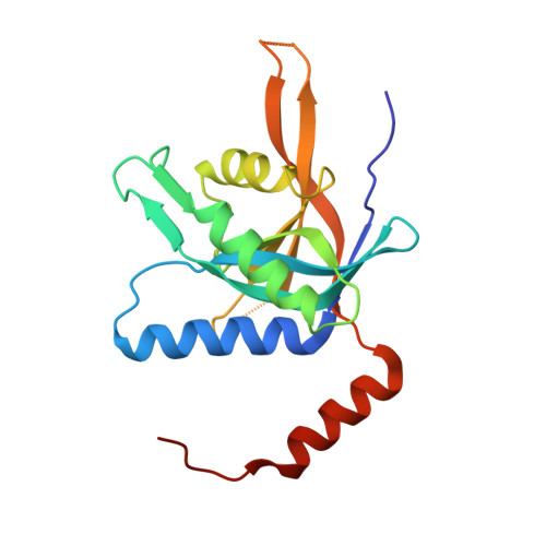
    The 26S proteasome is a large protein complex involved in protein degradation. We have shown previously that the PSMD7/Mov34 subunit of the human proteasome contains a proteolytically resistant MPN domain. MPN domain family members comprise subunits of the proteasome, COP9-signalosome and translation initiation factor 3 complexes. Here, the crystal structure of two C-terminally truncated proteins, MPN 1-186 and MPN 1-177, were solved to 1.96 and 3.0 A resolution, respectively. MPN 1-186 is formed by nine beta-strands surrounded by three alpha-helices plus a fourth alpha-helix at the C terminus. This final alpha-helix emerges from the domain core and folds along with a symmetrically related subunit, typical of a domain swap. The crystallographic dimer is consistent with size-exclusion chromatography and DLS analysis showing that MPN 1-186 is a dimer in solution. MPN 1-186 shows an overall architecture highly similar to the previously reported crystal structure of the Archaeal MPN domain AfJAMM of Archaeoglobus fulgidus. However, previous structural and biophysical analyses have shown that neither MPN 1-186 nor full-length human Mov34 bind metal, in opposition to the zinc-binding AfJAMM structures. The zinc ligand residues observed in AfJAMM are conserved in the yeast Rpn11 proteasome and Csn5 COP-signalosome subunits, which is consistent with the isopeptidase activity described for these proteins. The results presented here show that, although the MPN domain of Mov34 shows a typical metalloprotease fold, it is unable to coordinate a metal ion. This finding and amino acid sequence comparisons can explain why the MPN-containing proteins Mov34/PSMD7, RPN8, Csn6, Prp8p and the translation initiation factor 3 subunits f and h do not show catalytic isopeptidase activity, allowing us to propose the hypothesis that in these proteins the MPN domain has a primarily structural function.


  • Organizational Affiliation

    Center for Structural Molecular Biology, Brazilian Synchrotron Light Laboratory (LNLS), Campinas, SP, Brazil. msanches@lnls.br


Macromolecules
Find similar proteins by:  (by identity cutoff)  |  3D Structure
Entity ID: 1
MoleculeChains Sequence LengthOrganismDetailsImage
26S proteasome non-ATPase regulatory subunit 7
A, B
187Homo sapiensMutation(s): 0 
Gene Names: PSMD7Mov34L
UniProt & NIH Common Fund Data Resources
Find proteins for P51665 (Homo sapiens)
Explore P51665 
Go to UniProtKB:  P51665
PHAROS:  P51665
GTEx:  ENSG00000103035 
Entity Groups  
Sequence Clusters30% Identity50% Identity70% Identity90% Identity95% Identity100% Identity
UniProt GroupP51665
Sequence Annotations
Expand
  • Reference Sequence
Small Molecules
Ligands 5 Unique
IDChains Name / Formula / InChI Key2D Diagram3D Interactions
12P
Query on 12P

Download Ideal Coordinates CCD File 
D [auth A]DODECAETHYLENE GLYCOL
C24 H50 O13
WRZXKWFJEFFURH-UHFFFAOYSA-N
ETE
Query on ETE

Download Ideal Coordinates CCD File 
J [auth B]2-{2-[2-2-(METHOXY-ETHOXY)-ETHOXY]-ETHOXY}-ETHANOL
C9 H20 O5
ZNYRFEPBTVGZDN-UHFFFAOYSA-N
PG4
Query on PG4

Download Ideal Coordinates CCD File 
E [auth A]TETRAETHYLENE GLYCOL
C8 H18 O5
UWHCKJMYHZGTIT-UHFFFAOYSA-N
PGE
Query on PGE

Download Ideal Coordinates CCD File 
F [auth A],
G [auth A],
K [auth B],
L [auth B]
TRIETHYLENE GLYCOL
C6 H14 O4
ZIBGPFATKBEMQZ-UHFFFAOYSA-N
SO4
Query on SO4

Download Ideal Coordinates CCD File 
C [auth A],
H [auth B],
I [auth B]
SULFATE ION
O4 S
QAOWNCQODCNURD-UHFFFAOYSA-L
Experimental Data & Validation

Experimental Data

  • Method: X-RAY DIFFRACTION
  • Resolution: 1.95 Å
  • R-Value Free: 0.240 
  • R-Value Work: 0.201 
  • R-Value Observed: 0.203 
  • Space Group: C 1 2 1
Unit Cell:
Length ( Å )Angle ( ˚ )
a = 114.103α = 90
b = 95.289β = 121.13
c = 58.988γ = 90
Software Package:
Software NamePurpose
SCALAdata scaling
SHARPphasing
SOLOMONphasing
REFMACrefinement
PDB_EXTRACTdata extraction
MAR345dtbdata collection
MOSFLMdata reduction

Structure Validation

View Full Validation Report


Entry History 

Deposition Data

Revision History  (Full details and data files)

  • Version 1.0: 2007-07-17
    Type: Initial release
  • Version 1.1: 2008-05-01
    Changes: Version format compliance
  • Version 1.2: 2011-07-13
    Changes: Derived calculations, Version format compliance
  • Version 1.3: 2023-12-27
    Changes: Data collection, Database references, Derived calculations, Refinement description