Structural and functional relationships in the virulence-associated cathepsin L proteases of the parasitic liver fluke, Fasciola hepatica.
Stack, C.M., Caffrey, C.R., Donnelly, S.M., Seshaadri, A., Lowther, J., Tort, J.F., Collins, P.R., Robinson, M.W., Xu, W., McKerrow, J.H., Craik, C.S., Geiger, S.R., Marion, R., Brinen, L.S., Dalton, J.P.(2008) J Biol Chem 283: 9896-9908
- PubMed: 18160404
- DOI: https://doi.org/10.1074/jbc.M708521200
- Primary Citation of Related Structures:
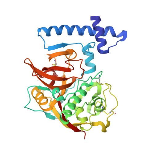
2O6X - PubMed Abstract:
The helminth parasite Fasciola hepatica secretes cysteine proteases to facilitate tissue invasion, migration, and development within the mammalian host. The major proteases cathepsin L1 (FheCL1) and cathepsin L2 (FheCL2) were recombinantly produced and biochemically characterized. By using site-directed mutagenesis, we show that residues at position 67 and 205, which lie within the S2 pocket of the active site, are critical in determining the substrate and inhibitor specificity. FheCL1 exhibits a broader specificity and a higher substrate turnover rate compared with FheCL2. However, FheCL2 can efficiently cleave substrates with a Pro in the P2 position and degrade collagen within the triple helices at physiological pH, an activity that among cysteine proteases has only been reported for human cathepsin K. The 1.4-A three-dimensional structure of the FheCL1 was determined by x-ray crystallography, and the three-dimensional structure of FheCL2 was constructed via homology-based modeling. Analysis and comparison of these structures and our biochemical data with those of human cathepsins L and K provided an interpretation of the substrate-recognition mechanisms of these major parasite proteases. Furthermore, our studies suggest that a configuration involving residue 67 and the "gatekeeper" residues 157 and 158 situated at the entrance of the active site pocket create a topology that endows FheCL2 with its unusual collagenolytic activity. The emergence of a specialized collagenolytic function in Fasciola likely contributes to the success of this tissue-invasive parasite.
Organizational Affiliation:
Institute for the Biotechnology of Infectious Diseases, University of Technology Sydney, Sydney, New South Wales 2007, Australia.