2O4U

Crystal structure of Mammalian Dimeric Dihydrodiol Dehydrogenase


Experimental Data Snapshot

  • Method: X-RAY DIFFRACTION
  • Resolution: 2.00 Å
  • R-Value Free: 0.193 
  • R-Value Work: 0.161 
  • R-Value Observed: 0.163 

wwPDB Validation   3D Report Full Report


This is version 1.2 of the entry. See complete history


Literature

Structures of dimeric dihydrodiol dehydrogenase apoenzyme and inhibitor complex: probing the subunit interface with site-directed mutagenesis.

Carbone, V.Endo, S.Sumii, R.Chung, R.P.Matsunaga, T.Hara, A.El-Kabbani, O.

(2008) Proteins 70: 176-187

  • DOI: https://doi.org/10.1002/prot.21566
  • Primary Citation of Related Structures:  
    2O48, 2O4U

  • PubMed Abstract: 

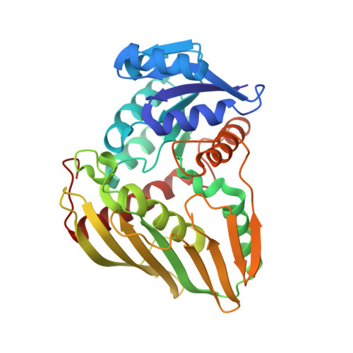
    Dimeric dihydrodiol dehydrogenase (DD) catalyses the nicotinamide adenine dinucleotide phosphate (NADP+)-dependent oxidation of trans-dihydrodiols of aromatic hydrocarbons to their corresponding catechols. This is the first report of the crystal structure of the dimeric enzyme determined at 2.0 A resolution. The tertiary structure is formed by a classical dinucleotide binding fold comprising of two betaalphabetaalphabeta motifs at the N-terminus and an eight-stranded, predominantly antiparallel beta-sheet at the C-terminus. The active-site of DD, occupied either by a glycerol molecule or the inhibitor 4-hydroxyacetophenone, is located in the C-terminal domain of the protein and maintained by a number of residues including Lys97, Trp125, Phe154, Leu158, Val161, Asp176, Leu177, Tyr180, Trp254, Phe279, and Asp280. The dimer interface is stabilized by a large number of intermolecular contacts mediated by the beta-sheet of each monomer, which includes an intricate hydrogen bonding network maintained in principal by Arg148 and Arg202. Site-directed mutagenesis has demonstrated that the intact dimer is not essential for catalytic activity. The similarity between the quaternary structures of mammalian DD and glucose-fructose oxidoreductase isolated from the prokaryotic organism Zymomonas mobilis suggests that both enzymes are members of a unique family of oligomeric proteins and may share a common ancestral gene.


  • Organizational Affiliation

    Department of Medicinal Chemistry, Victorian College of Pharmacy, Monash University, Parkville, Victoria 3052, Australia.


Macromolecules
Find similar proteins by:  (by identity cutoff)  |  3D Structure
Entity ID: 1
MoleculeChains Sequence LengthOrganismDetailsImage
Dimeric dihydrodiol dehydrogenaseA [auth X]334Macaca fascicularisMutation(s): 0 
EC: 1.3.1.20
UniProt
Find proteins for Q9TQS6 (Macaca fascicularis)
Explore Q9TQS6 
Go to UniProtKB:  Q9TQS6
Entity Groups  
Sequence Clusters30% Identity50% Identity70% Identity90% Identity95% Identity100% Identity
UniProt GroupQ9TQS6
Sequence Annotations
Expand
  • Reference Sequence
Small Molecules
Ligands 3 Unique
IDChains Name / Formula / InChI Key2D Diagram3D Interactions
PO4
Query on PO4

Download Ideal Coordinates CCD File 
B [auth X]
C [auth X]
D [auth X]
E [auth X]
F [auth X]
B [auth X],
C [auth X],
D [auth X],
E [auth X],
F [auth X],
G [auth X],
H [auth X]
PHOSPHATE ION
O4 P
NBIIXXVUZAFLBC-UHFFFAOYSA-K
GOL
Query on GOL

Download Ideal Coordinates CCD File 
J [auth X]GLYCEROL
C3 H8 O3
PEDCQBHIVMGVHV-UHFFFAOYSA-N
BME
Query on BME

Download Ideal Coordinates CCD File 
I [auth X]BETA-MERCAPTOETHANOL
C2 H6 O S
DGVVWUTYPXICAM-UHFFFAOYSA-N
Experimental Data & Validation

Experimental Data

  • Method: X-RAY DIFFRACTION
  • Resolution: 2.00 Å
  • R-Value Free: 0.193 
  • R-Value Work: 0.161 
  • R-Value Observed: 0.163 
  • Space Group: P 61 2 2
Unit Cell:
Length ( Å )Angle ( ˚ )
a = 123.177α = 90
b = 123.177β = 90
c = 121.127γ = 120
Software Package:
Software NamePurpose
REFMACrefinement
HKL-2000data collection
HKL-2000data reduction
SCALEPACKdata scaling
Cootmodel building

Structure Validation

View Full Validation Report


Entry History 

Deposition Data

Revision History  (Full details and data files)

  • Version 1.0: 2007-07-31
    Type: Initial release
  • Version 1.1: 2011-07-13
    Changes: Derived calculations, Version format compliance
  • Version 1.2: 2023-10-25
    Changes: Data collection, Database references, Derived calculations, Refinement description